Cleft Palate with Cleft Lip
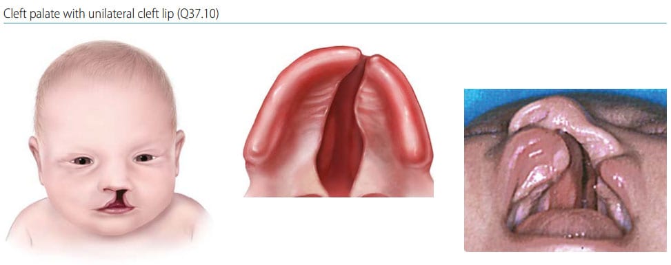
Cleft palate with cleft lip is characterized as a cleft of the upper lip extending through the hard palate (primary and secondary palate), and might also extend through the soft palate (see Fig. 27).
Fig. 27. Cleft palate with cleft lip

Photograph source: Dr Pedro Santiago and Dr Miguel Yáñez (EE. UU.).
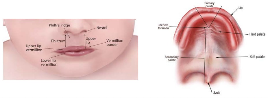
Fig. 28. Anatomy of the lip and palate

Diagnosis
Prenatal. It can be suspected prenatally but can easily be missed or misdiagnosed. Cases identified or suspected prenatally should be confirmed postnatally before inclusion in a surveillance programme.
Postnatal. Cleft lip with cleft palate is easily recognized on physical examination after delivery, provided the palate is also checked carefully.
Clinical and epidemiologic notes
Rarer conditions that can be confused with typical cleft lip are the atypical or Tessier type clefts and the amniotic band spectrum.
- Atypical or Tessier type craniofacial clefts are a group of clefting defects that involve the cranial and/or facial skeleton. The 14 various Tessier’s clefts extend through radiating axes of facial and cranial bones, including towards the eye or nasolacrimal canal, or even more laterally towards the ear. Amniotic band spectrum can cause facial disruptions that involve both the lip and palate, and often include atypical skull and brain lesions (e.g. atypical encephaloceles).
Cleft lip with cleft palate can be associated with other birth defects or syndromes, more commonly so than cleft lip alone.
Additional clinical tips:
- Always check the palate when you see a cleft lip – the diagnosis is important both for surveillance and for clinical care.
- Check for lip pits in the lower lip (Fig. 23), in the child and in the parents – it is a sign of a genetic condition (van der Woude syndrome) with high recurrence risk (a parent may have the pits but not the cleft).
- When in doubt that what you see is a typical cleft, take a photograph or a very careful drawing – such documentation helps the clinical reviewer reach a correct diagnosis.
Checklist for high-quality reporting
| Cleft Lip with Cleft Palate – Documentation Checklist |
Describe in detail, including:
Describe procedures to assess further additional malformations, and if present describe. Take and report photographs: Very useful; can be crucial for review. Report whether specialty consultation(s) were done, and if so, report the results. Plastic surgery consultation reports are often useful. |
Key visuals:
Anatomy of the lip and palate. Note that in bilateral cleft lip, a median remnant of the philtrum is still present (Fig. 28). Note also that the clefting can extend to the gum or alveolus but does not extend beyond the incisive foramen (thus involving only the primary palate).