Large Intestinal Atresia/Stenosis
Large intestinal atresia or stenosis – also known as colonic atresia – is the complete or partial obstruction of the opening (lumen) within the colon.
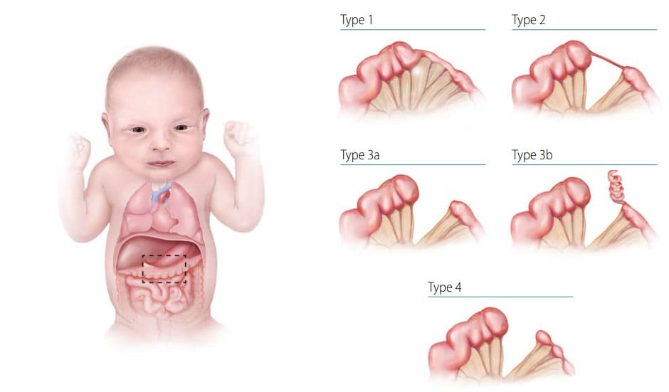
Fig. 30. Main types of large intestinal atresia

- Type: Four types of atresia have been recognized (see Fig. 30):
- In type 1, the lumen is occluded by internal tissue (mucosal web) with intact mesentery.
- In type 2, the atretic segment is a fibrous cord, and connects the two ends of the large intestine.
- In type 3a, the atretic segment occurs with a V-shaped mesenteric gap defect (in type 3b, there is an “apple peel” appearance of a portion of the gut).
- In type 4, the atresia involves two or more regions of the colon, with an appearance described as a string of sausages (approximately one third of all large intestinal atresias are type 4).
- Location: Sigmoid and transverse regions are most commonly affected.
Diagnosis
Prenatal. These conditions are difficult to detect and diagnose prenatally, so any prenatal diagnosis should be confirmed postnatally.
Postnatal. Large intestinal atresia or stenosis should be suspected in the newborn infant who fails to pass meconium or stool, has abdominal distention and/or bilious vomiting. Diagnosis is confirmed through direct imaging of the bowel by x-ray, barium enema, surgery, or autopsy. Partial colonic stenosis may be diagnosed later, even weeks after delivery.
Clinical and epidemiologic notes
Large intestinal atresia is rare, with an estimated birth prevalence of approximately 1 in 20 000 births. This condition represents less than 10% of all intestinal atresias.
Checklist for high-quality reporting
| Large Intestinal Atresia/Stenosis – Documentation Checklist |
Describe the anatomy in detail, best using a combination of clinical, imaging, and surgical reports; specifically include information on the following elements:
Describe procedures to assess further additional malformations:
Copy and attach key imaging. Report findings of surgery or autopsy. Report whether specialty consultation(s) were done and if so, report the results. |