Interrupted Aortic Arch
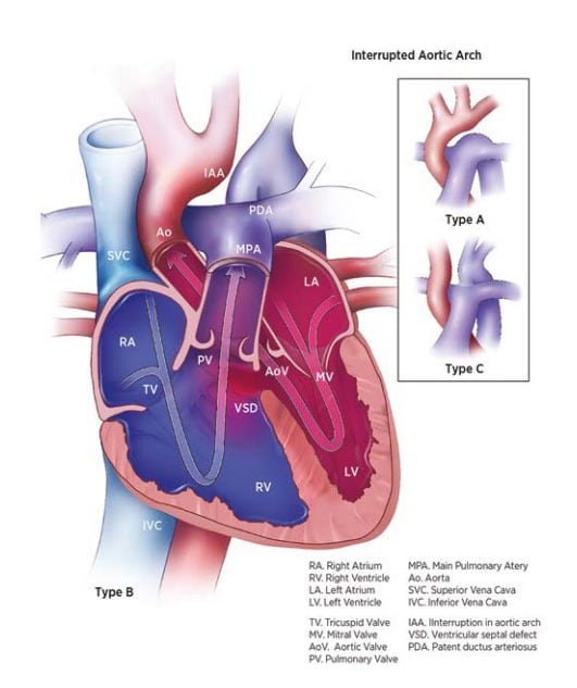
Interrupted aortic arch (IAA) is a structural heart defect characterized anatomically by a discontinuity (interruption) along the aortic arch. Depending on the site of discontinuity, IAA is classified into three types (see Fig. 20), of which type B is the most frequent (50–70%). Type A is less common (30–45%) and type C is rare.
- Type A: The discontinuity is distal to the left subclavian artery (approximately in the same region as coarctation of the aorta).
- Type B, the most common form: The discontinuity is more proximal, between the left carotid and subclavian.
- Type C: The discontinuity is more proximal still, between the brachiocephalic artery and the common carotid artery.
Fig. 20. Interrupted aortic arch


Diagnosis
Prenatal. IAA is easily missed on the obstetric anomaly scan, though it might be suspected based on discrepancy between the left and right ventricular sizes. Whereas in expert hands fetal echocardiography can provide a firm diagnosis, prenatally diagnosed cases should be confirmed postnatally.
Postnatal. Infants can present clinically in the early neonatal period, when the ductus closes, with signs and symptoms of congestive heart failure and systemic hypoperfusion (cardiogenic shock). Newborn screening via pulse oximetry can lead to earlier diagnosis.
Clinical and epidemiologic notes
As noted, early presentation is one of heart failure and cardiogenic shock, with rapid clinical deterioration as the ductus closes. Because of right-to-left shunting at the ductus arteriosus, infants may initially show differential oxygen saturation or cyanosis. In type A, the difference in saturation is between upper and lower limbs (the latter being lower); in type B, it is between the left and right arm (lower saturation in the left).
Ventricular septal defect and other intracardiac defects are often present.
The three types of IAA differ in their association with genetic risk factors. For example, deletion 22q11 occurs in 50% or more of cases of type B IAA, and is rare in the other types. Other syndromes that can occur with IAA include CHARGE syndrome (Q30.01).
Checklist for high-quality reporting
| Interrupted Aortic Arch (IAA) – Documentation Checklist |
Describe in detail the clinical and echocardiographic findings:
Look for and document extracardiac birth defects: IAA can occur with genetic syndromes such as deletion 22q11, which is associated with many external and internal anomalies. Report whether specialty consultation(s) was done (e.g. a paediatric cardiologist or geneticist). Report any genetic testing and results (e.g. chromosomal studies, genomic microarray, etc.). |