Renal Agenesis/Hypoplasia
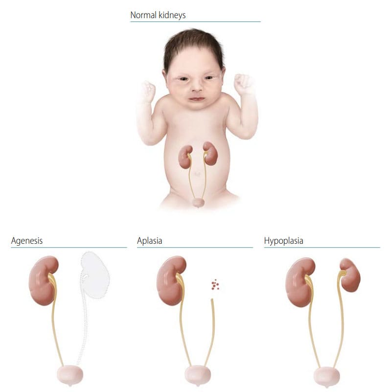
Renal agenesis is a complete absence of one (unilateral) or both (bilateral) kidneys, whereas in renal aplasia the kidney has failed to develop beyond its most primitive form. In practice, renal agenesis and renal aplasia might be indistinguishable. Renal hypoplasia is a congenitally small kidney without dysplasia and can be bilateral or unilateral (see Fig. 33).
Fig. 33. Renal agenesis/hypoplasia

- Location: Unilateral or bilateral; if bilateral, findings can be asymmetric (e.g. agenesis on one side, hypoplasia on the other side).
- Agenesis/degree of hypoplasia: Kidney absent versus present but small or rudimentary.
Note: Be sure not to confuse this condition with multicystic dysplastic kidney or multicystic renal dysplasia.
Diagnosis
Prenatal. Renal agenesis can be diagnosed or strongly suspected prenatally by ultrasound but should always be confirmed postnatally.
Postnatal. Renal agenesis or hypoplasia is conclusively diagnosed only through direct assessment by abdominal ultrasound, CT or MRI scan, surgery, or autopsy. Bilateral renal agenesis should be considered in an infant with features of Potter sequence. Bilateral renal hypoplasia might or might not be recognized after delivery, depending on the severity and degree of residual kidney function. Unilateral renal agenesis or hypoplasia may be clinically silent at delivery if the contralateral kidney is functional, such that the diagnosis may occur months or years after birth (if at all).
Clinical and epidemiologic notes
In about half of all cases of bilateral renal agenesis there are other structural anomalies (e.g. urogenital, cardiac, skeletal, central nervous system) or syndromes (chromosomal or genetic).
- VATER/VACTERL (vertebral, anus, cardiac, trachea, oesophagus, renal, limb) association;
- MURCS association (Mullerian, renal, cervicothoracic somite abnormalities), a developmental disorder affecting primarily females and involving mainly the reproductive and urinary systems;
- sirenomelia; and
- caudal dysplasia (also seen in pregestational diabetes).
- Bilateral renal agenesis is a lethal condition – the fetus may be stillborn or die shortly after delivery.
- Look for major anomalies and minor anomalies – renal agenesis is seen in hundreds of genetic conditions, including common trisomies, deletion 22q11, Melnick-Fraser syndrome, Fraser cryptophthalmos syndrome, and branchio-oto-renal syndrome.
- Determine related anomalies in bilateral renal agenesis (Potter sequence: abnormal facies, talipes [clubfoot] and other contractures, pulmonary hypoplasia).
- Distinguish renal agenesis from other kidney anomalies (multicystic dysplasia and polycystic renal disease).
Checklist for high-quality reporting
| Renal Agenesis/Hypoplasia – Documentation Checklist |
Describe in detail; including:
Take and report photographs of any external defects: Especially show clearly the location of the urethra; can be crucial for review. Describe evaluations to find or rule out related and associated anomalies:
Report whether autopsy (pathology) findings are available and if so, report the results. |