4.8 Congenital Malformations of Genital Organs Hypospadias (Q54.0–Q54.9)
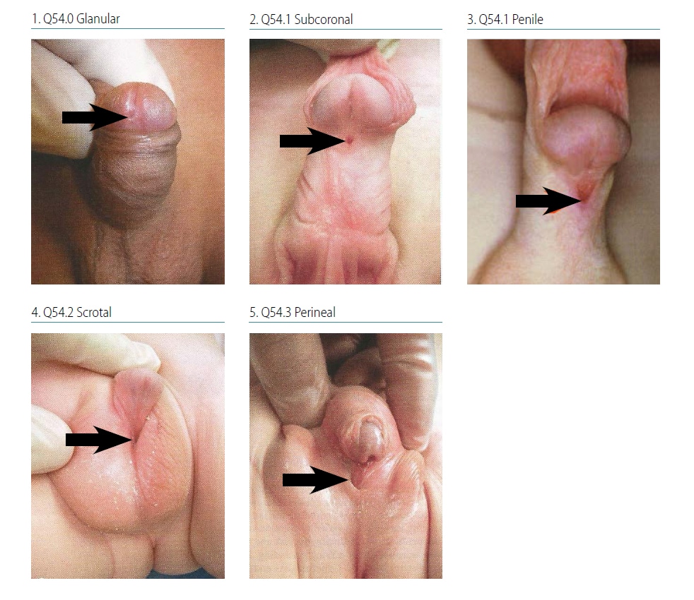
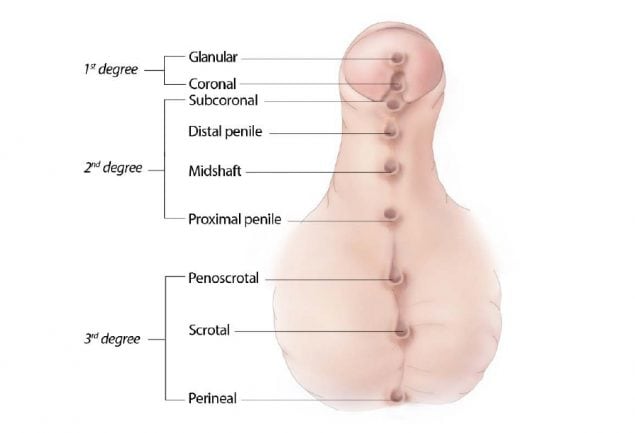
Hypospadias is characterized by an abnormal (ventral) placement of the external urethral meatus in male infants. Normal placement of the urethral meatus is on the tip of the penis, whereas in hypospadias the meatus is ventrally and proximally displaced (on the underside of the penis). Hypospadias is classified by severity (see Fig. 4.31), depending on the location of the meatus: first-degree hypospadias includes the more distal forms, glanular and coronal; second-degree hypospadias includes subcoronal and penile shaft hypospadias; and third-degree – the most severe – includes scrotal and perineal hypospadias.
The shortening of the ventral side of the penis found in hypospadias can result in a curvature of the penis, known as chordee. Chordee is common in severe cases of hypospadias, but it can also occur independently. Chordee by itself is considered a minor anomaly.

Photograph source: 1, 2, 4, 5: Nelson Textbook of Pediatrics (http://www.slideshare.net/wadoodaref/congenital-anomalies-videosession-v3-11680674external icon).
Relevant ICD-10 codes
Q54.0 Hypospadias, balanic (coronal, glanular)
Q54.1 Hypospadias, penile (includes subcoronal and midshaft)
Q54.2 Hypospadias, penoscrotal
Q54.3 Hypospadias, perineal
Q54.8 Hypospadias, other
Q54.9 Hypospadias, unspecified
Diagnosis
Prenatal. Hypospadias is difficult to diagnose prenatally by ultrasonography, and might be confused with micropenis, penile cyst, chordee or ambiguous genitalia. For this reason, a prenatal diagnosis of hypospadias should always be confirmed postnatally. When this is not possible (e.g. termination of pregnancy or unexamined fetal death), the programme should have criteria in place to determine whether to accept or not accept a case based solely on prenatal data.
Postnatal. A careful, systematic examination of the male newborn should allow a firm diagnosis of hypospadias. Note that the milder forms such as balanic (glanular) hypospadias are easily missed at delivery and might be discovered during circumcision. The surgical report might provide definitive detail of the urethral placement and whether chordee are present.
Clinical and epidemiologic notes
The diagnosis of hypospadias might be missed, especially with the less severe forms. Observing the flow of urine can aid in the diagnosis and in establishing the degree/severity of the condition. Identifying severity is important because of the different disease associations and the clinical impact. The most severe forms of hypospadias (e.g. scrotal hypospadias) are more often associated with syndromes compared to the milder forms (e.g. balanic [glanular]) of hypospadias.
Hypospadias is often an isolated (> 80%), non-syndromic anomaly. However, the proportion of isolated defects decreases with increasing severity (penile and scrotal) of hypospadias. For this reason, it is crucial to report all findings. Obtaining good clinical photographs is important for the expert reviewer but is not easy to do. Documenting the severity using drawing might help in selected cases.
Suggested non-genetic risk factors for hypospadias include advanced maternal age (> 35 years) and environmental exposure to certain chemicals (endocrine disruptors).
The birth prevalence of hypospadias varies widely, between 2 and 39 per 10 000 births.
Inclusions
Q54.0 Hypospadias, balanic (coronal, glanular)
Q54.1 Hypospadias, penile (includes subcoronal and midshaft)
Q54.2 Hypospadias, penoscrotal
Q54.3 Hypospadias, perineal
Q54.8 Hypospadias, other
Q54.9 Hypospadias, unspecified
Exclusions
Q54.4 Chordee (a minor anomaly if isolated)
Checklist for high-quality reporting
| Hypospadias – Documentation Checklist |
Describe in detail:
Include photographs: Show clearly the location of the urethra or use drawing; can be crucial for review. Describe evaluations to find or rule out related and associated anomalies:
Report whether autopsy (pathology) findings are available and if so, report the results. |
Suggested data quality indicators
| Category | Suggested Practices and Quality Indicators |
| Description and documentation | Review sample for documentation of key descriptors – location of urethral meatus:
|
| Coding |
|
| Clinical classification |
|
| Prevalence |
|
| Key illustration |  Note: Illustration indicates all possible locations for the malformation, but typically a case will only have an opening in one location. |
RENAL AGENESIS/HYPOPLASIA (Q60.0–Q60.5)
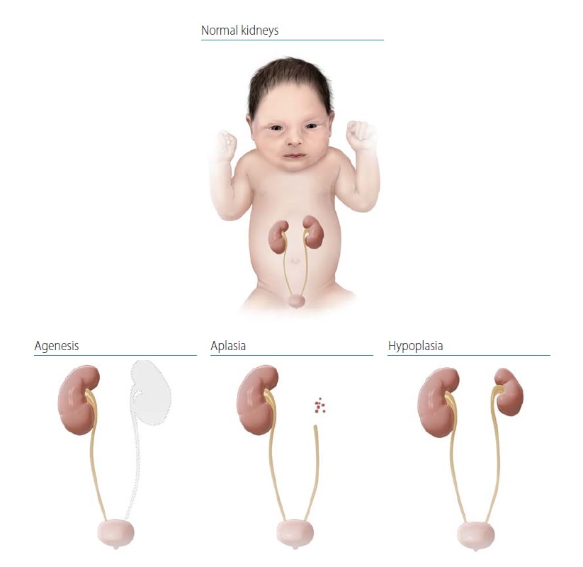
Renal agenesis is a complete absence of one (unilateral) or both (bilateral) kidneys. If bilateral, renal agenesis is a lethal condition: the fetus will be stillborn or die shortly after delivery. In utero, bilateral renal agenesis leads to oligohydramnios (too little amniotic fluid), which causes Potter sequence (syndrome) – abnormal facies, talipes (clubfoot) and other contractures, and pulmonary hypoplasia.
Renal hypoplasia is a congenitally small kidney (< 50% of expected weight) without dysplasia, which can be bilateral or unilateral. Renal hypoplasia might be associated with hydronephrosis but is not usually associated with abnormal ureters or other urinary tract findings.
Renal agenesis must be distinguished from renal multicystic dysplasia (Q61.40 and Q61.41) – considered a specific developmental anomaly – and from polycystic renal disease (Q61.1, Q61.2, Q61.3, a single-gene disorder with infantile and later-onset forms).

These have different etiologies and ICD codes.
Relevant ICD-10 codes
Q60.0 Unilateral renal agenesis
Q60.1 Bilateral renal agenesis
Q60.2 Unspecified renal agenesis
Q60.3 Renal hypoplasia, unilateral
Q60.4 Renal hypoplasia, bilateral
Q60.5 Renal hypoplasia, unspecified
Q60.6 Potter sequence with renal agenesis
Diagnosis
Prenatal. Bilateral renal agenesis or severe hypoplasia should be suspected prenatally in a pregnancy with severe oligohydramnios (urine from fetal kidneys accounts for most of the amniotic fluid from 14 weeks’ gestations onward). Fetal kidneys might be small or absent, and the bladder might not be visualized (empty). In contrast, multicystic/polycystic kidneys tend to be large and “bright” on fetal ultrasound. Of note, in some cases, large dysplastic kidneys can reduce and disappear by the time of birth, and in others a kidney might be absent on one side and dysplastic on the contralateral side, suggesting that some cases of agenesis might have started as dysplasia.
Postnatal. At delivery, bilateral renal agenesis should be considered in an infant with features of Potter sequence (Q60.6), which include respiratory distress (due to pulmonary hypoplasia), characteristic facial traits (wide-set eyes, flat face, low-set large ears, small chin, loose or excessive skin), and joint contractures (talipes and others). Bilateral renal hypoplasia might or might not be recognized after delivery, depending on the severity and degree of residual kidney function. Renal agenesis or hypoplasia is conclusively diagnosed only through direct assessment by abdominal ultrasound, CT or MRI scan, surgery or autopsy.
Unilateral renal agenesis or hypoplasia can be clinically silent at delivery if the contralateral kidney is not impaired, such that the diagnosis might occur months or years after birth, if at all. Some unilateral cases are diagnosed only as incidental findings during evaluation for other conditions, or in screening of family members of a patient with bilateral renal agenesis.
Clinical and epidemiologic notes
At least half of cases of bilateral renal agenesis are estimated to be associated with other structural anomalies (e.g. urogenital, cardiac, skeletal, CNS) or syndromes (chromosomal or genetic). The non-syndromic multiple anomaly patterns include the VATER/ VACTERL association (vertebral, anus, cardiac, trachea, oesophagus, renal, limb (radial agenesis); Q87.26), MURCS (mulllerian, renal cervicothoracic, somite) association (Q51.8), sirenomelia (Q87.24), and caudal dysplasia “syndrome” (also seen in maternal pregestational diabetes). Renal agenesis is seen in hundreds of genetic conditions (Mendelian and chromosomal), including common trisomies, deletion 22q11, Melnick-Fraser syndrome, Fraser cryptophthalmos syndrome and branchio-oto-renal syndrome.
Bilateral renal agenesis occurs in 1 in 4000 births and is more common in stillbirths and in males. Unilateral renal agenesis is more common on the left side, is associated with an absent ureter on the same side, and at times with renal hypoplasia in the contralateral kidney. The frequency of unilateral agenesis is estimated as 1 in 3000 births but it is likely underdiagnosed. Nongenetic risk factors include maternal pregestational diabetes.
Inclusions
Q60.0 Unilateral renal agenesis
Q60.1 Bilateral renal agenesis
Q60.3 Renal hypoplasia, unilateral
Q60.4 Renal hypoplasia, bilateral
Unilateral renal agenesis with contralateral renal hypoplasia
Renal agenesis is a defect reported as part of VATER or VACTERL association (vertebral, anus, cardiac, trachea, oesophagus, renal, limb (radial agenesis)).
Exclusions
Q61.1, Q61.19 Autosomal recessive, polycystic kidney, infantile type
Q61.11-Q61.3 Multicystic dysplastic kidney, multicystic renal dysplasia
Checklist for high-quality reporting
| Renal Agenesis/Hypoplasia – Documentation Checklist |
Describe in detail, including:
Take and report photographs of any external defects: Especially show clearly the location of the urethra; can be Describe evaluations to find or rule out related and associated anomalies:
Report whether autopsy (pathology) findings are available and if so, report the results. |
Suggested data quality indicators
| Category | Suggested Practices and Quality Indicators |
| Description and documentation |
|
| Coding |
|
| Clinical classification |
|
| Prevalence |
|