Congenital Anomalies and Deformations of the Musculoskeletal System: Talipes Equinovarus
Talipes equinovarus (TEV) is the specific term and common type of what is sometimes called “clubfoot”, a term that encompasses a range of anomalies of the ankle or foot present at birth.
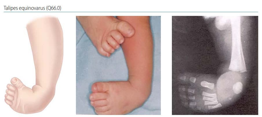
Fig. 34. Talipes equinovarus

Photograph and x-ray source: Dr Idalina Montes and Dr Rafael Longo (Puerto Rico)
Key findings in TEV (see Fig. 34):
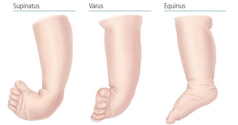
- Position: Fixation of the foot (forefoot and hindfoot) in plantar flexion (equinus), deviation toward the midline (varus) and upward rotation so the foot rests on its outer side (supinatus).
- Severity: Milder cases are “positional”, meaning that the foot and ankle can be gently manipulated into a normal position. In more severe cases the foot and ankle can be “rigid” or ‘”fixed”, in that they cannot be manipulated into a normal position and require orthopaedic or surgical treatment.
Diagnosis
Prenatal. Clubfoot can be identified or suspected on prenatal ultrasound. However, it should not be included in birth defects surveillance data without postnatal confirmation.
Postnatal. Clubfoot is readily diagnosed in the newborn examination. Cases should be followed and evaluated sequentially to assess the degree of severity and whether treatment other than manipulation is necessary.
Clinical and epidemiologic notes
- Milder cases are “positional”, meaning that the foot can be gently manipulated into a normal position and typically does not require orthopaedic or surgical interventions. Positional talipes is excluded in surveillance.
- More severe cases are “rigid” or “fixed”, meaning the foot cannot be manipulated into a normal position and requires orthopaedic or surgical treatment.
Imaging is very helpful but a clinical examination in expert hands can provide a firm diagnosis.
- TEV can occur with deformations in other joints (e.g. elbow, hands, knees) – check and report.
- TEV can be due to many syndromes and disorders, especially of the brain and bone/cartilage – evaluate carefully.
- Be sure to differentiate TEV from other types of “clubfoot”, such as talipes calcaneovalgus, common in trisomy 18 (in which the ankle joint is dorsiflexed instead of plantar flexed, and the forefoot deviated outwards); and talipes calcaneovarus (in which the ankle joint is dorsiflexed, and the forefoot deviated inwards).
Checklist for high-quality reporting
| Talipes Equinovarus – Documentation Checklist |
Describe in detail, including:
Describe other deformations if present (e.g. knees, fingers, elbows), especially if rigid (e.g. in arthrogryposis). Describe procedures to assess further additional malformations and, if one or more is present, describe. Take and report photographs of anomaly and whole baby; useful for review but not sufficient as confirmation. Specialty consultations: Report which were done (including genetics and orthopaedics) and results. Note that the following conditions are usually not considered eligible conditions:
Additional visual: foot positions 
|