4.5f Tricuspid Valve Atresia (Q22.4)
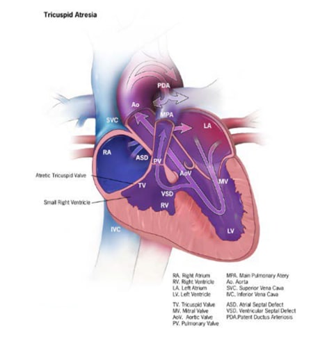
Tricuspid valve atresia is a structural heart defect characterized anatomically by a complete agenesis (failure of formation) of the tricuspid valve, leading to absence of direct communication and blood flow from the right atrium to the right ventricle. Having an atrial septal defect (ASD) (Fig. 4.19) is crucial for survival. Blood mixing causes significant cyanosis.
Fig. 4.19. Tricuspid valve atresia


Relevant ICD-10 codes
Q22.4 Tricuspid valve atresia (this code also include both atresia and stenosis)
Diagnosis
Prenatal. Tricuspid valve atresia can be readily suspected prenatally on a second trimester obstetric anatomic scan based on the absence of the tricuspid valve and the discrepancy in size of the ventricles (left ventricle > right ventricle). A suspected case should be confirmed postnatally.
Postnatal. The common clinical presentation in the newborn is cyanosis. Echocardiography has largely superseded other imaging techniques, although these have a role (e.g. catheterization to assess right ventricular pressures and resistance).
Newborn screening via pulse oximetry – which is based on the detection of low blood oxygen saturation – is expected to detect most cases of hypoxia due to tricuspid atresia.
Clinical and epidemiologic notes
Survival of the newborn depends on the presence of an ASD to allow the exit of blood from the right atrium to the left atrium (see Fig. 4.19), and an open ductus arteriosus to allow blood to reach the lungs to be oxygenated. Infants with only a small ASD or with a closing ductus will present early with severe symptoms.
Tricuspid atresia can co-occur with complex cardiovascular anomalies; for example, with heterotaxy, DORV or malposed great arteries. When the ventricular septum is intact, severe pulmonary valve stenosis or atresia might also be present, together with underdevelopment of the right ventricle.
Tricuspid atresia has been diagnosed in infants with deletion 22q11 (5–10%), common trisomies and other rarer genetic conditions.
Tricuspid atresia is one of the more common cyanotic CHDs, with a frequency of approximately 1 in 10 000 to 15 000 births. Tricuspid atresia is more common in males.
Inclusions
Q22.4 Tricuspid valve atresia (this code also include stenosis; see note below)
Notes:
- Although clinically severe tricuspid stenosis can resemble tricuspid atresia, most cases of tricuspid stenosis are mild. Tricuspid stenosis and tricuspid atresia should be kept separate in public health surveillance of prevalence and outcomes.
- Unfortunately, ICD-9 (International Classification of Diseases, Ninth Revision), ICD-10 and RCPCH coding systems use the same code for tricuspid atresia and stenosis. An option for differentiating the two conditions includes adding an additional digit to the ICD-10 core.
Exclusions
A programme must be clear about what is included when collecting data on tricuspid atresia and then apply the codes consistently.
Checklist for high-quality reporting
| Tricuspid Atresia – Documentation Checklist |
Describe in detail the clinical and echocardiographic findings:
Look for and document extracardiac birth defects and genetic conditions, such as deletion 22q11. Report whether specialty consultation(s) was done: Report on whether the diagnosis was made by a paediatric cardiologist, and whether the patient was seen by a geneticist. Report any genetic testing and results(e.g. chromosomal studies, genomic microarray, genomic sequencing). |
Suggested data quality indicators
| Category | Suggested Practices and Quality Indicators |
| Description and documentation | Review sample of clinical descriptions for documentation of key elements:
|
| Coding |
|
| Clinical classification |
|
| Prevalence |
|