4.2c Iniencephaly (Q00.2)
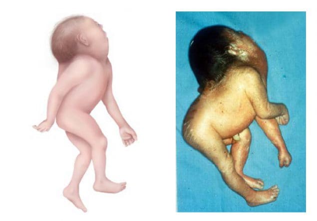
Iniencephaly is a rare and complex NTD involving the occiput and inion, resulting in extreme retroflexion of the head, variably combined with occipital encephalocele or rachischisis of the cervical or thoracic spine. The cranium is typically closed and skin covered, and an occipital encephalocele can be present. The face appears as upward looking; the neck, because of spine anomalies, is very short and might appear to be missing (see Fig 4.5). The occipital bone is abnormal leading to an enlarged foramen magnum with fusion of the cervical and thoracic vertebrae. The internal spine abnormalities result in the skin of the face being directly connected to the chest skin and the skin of the scalp continuous with the skin of the back. Many affected fetuses end in a miscarriage or are stillborn.
Fig. 4.5. Iniencephaly

Photograph source: CDC-Beijing Medical University collaborative project.
Relevant ICD-10 codes
Q00.2 Iniencephaly
Diagnosis
Prenatal. Iniencephaly might be difficult to diagnose precisely by ultrasound in the prenatal period, as it can be confused with other defects involving the brain and spine – encephalocele or craniorachischisis, as well as teratomas, goiter, lymphangioma and some syndromes. For this reason, a prenatal diagnosis of iniencephaly should always be confirmed postnatally. When this is not possible (e.g. termination of pregnancy or unexamined fetal death), the programme should have criteria in place to determine whether to accept or not accept a case based solely on prenatal data.
Postnatal. Careful examination of the fetus or newborn can confirm the diagnosis of iniencephaly and distinguish it from the other rare anomalies that involve the brain, cranium and spine. In iniencephaly, the cranial vault is always closed and the neck might appear shortened or not present.
Clinical and epidemiologic notes
Distinguishing iniencephaly from other abnormalities of the brain and spinal cord is important because these conditions have different causes and associated anomalies. With careful examination and radiograph confirmation, the diagnosis of iniencephaly is usually straightforward; however, the presence of an accompanying encephalocele or spina bifida might be confusing in some instances. The majority (84%) of affected infants have additional anomalies: micrognathia, cleft lip and palate, cardiovascular disorders, diaphragmatic hernias, and gastrointestinal malformations have been reported. Chromosomal conditions associated with iniencephaly include trisomy 13 and 18, and monosomy X. However, no population-based studies have been conducted to understand the proportion of isolated versus syndromic cases or the associated anomalies. It is crucial to report all physical findings and obtain good clinical photographs for the expert reviewer. Iniencephaly is reported to occur more commonly in females. Iniencephaly is a uniformly fatal condition.
Observational studies of non-genetic risk factors for iniencephaly are challenging given its very low prevalence. Presumably, since iniencephaly is an NTD, the non-genetic risk factors should be similar to other NTDs – pregestational diabetes, obesity, and hyperthermia (e.g. fever) in early pregnancy, and folic acid insufficiency/deficiency. Adequate periconceptional use of folic acid (as a supplement or through fortification) might also prevent iniencephaly.
Few reports on NTD prevalence specifically mention iniencephaly. The prevalence of iniencephaly is reported to vary widely between 0.1 to 10 per 10 000 births. Iniencephaly is counted when calculating total NTD prevalence.
Inclusions
Q00.2 Iniencephaly
Exclusions
Q00.0 Anencephaly, acrania, acphaly
Q00.1 Craniorachischisis
Checklist for high-quality reporting
| Iniencephaly – Documentation Checklist |
Describe in detail:
Report whether autopsy (pathology) findings are available and if so, report the results. |
Suggested data quality indicators
| Category | Suggested Practices and Quality indicators |
| Description and documentation |
|
| Coding |
|
| Clinical classification |
|
| Prevalence |
|