Longitudinal Preaxial (Tibia, Radius, First Ray)
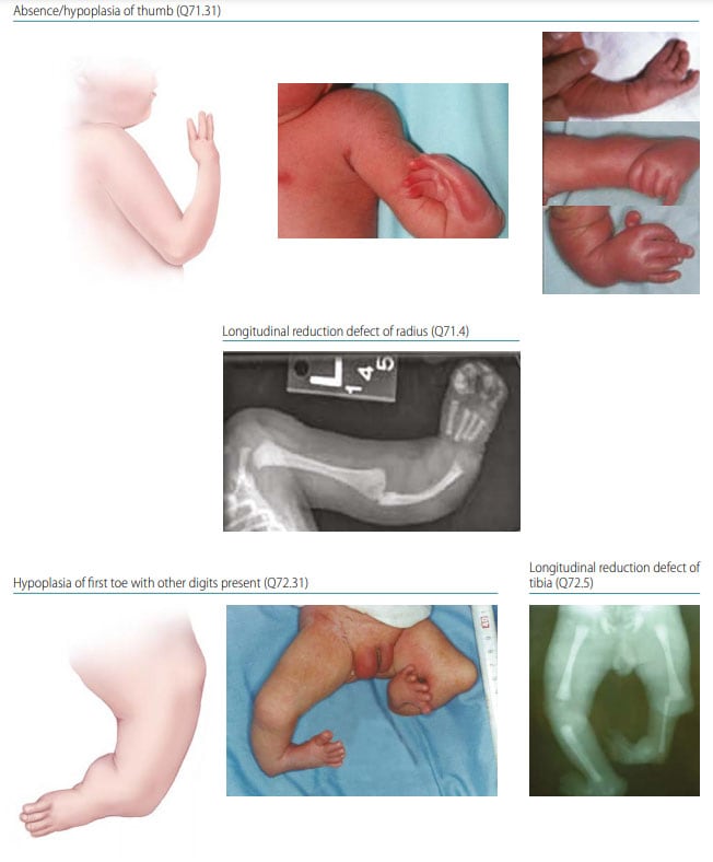
Fig. 42. Longitudinal preaxial

Photograph source: ECLAMC.
Preaxial limb deficiency is characterized by the absence or hypoplasia of the “preaxial” segments (those on the side of the thumb or big toe side) of the upper or lower limb (see Fig. 42). Preaxial limb deficiencies include:
- Hypoplasia/absence of the thumb (sometimes of the second finger)
- Hypoplasia/absence of the radius
- Hypoplasia/absence of the big toe (sometimes of the second toe)
- Hypoplasia/absence of the tibia.
Diagnosis
Prenatal. Preaxial limb deficiencies can be suspected prenatally but can easily be missed or misdiagnosed. Cases identified or suspected prenatally should be confirmed postnatally before inclusion in a surveillance programme.
Postnatal. The newborn examination can identify a longitudinal preaxial limb deficiency and distinguish it from other limb reduction defects (e.g. longitudinal postaxial defects). An accurate and complete diagnosis requires a detailed physical examination aided by radiography to characterize completely the bony anatomy.
Clinical and epidemiologic notes
Radial deficiency is often associated with hypoplasia or aplasia of the thumb and bowing of the ulna (so-called radial club hand, angulated to the radial side of the wrist). Isolated thumb hypoplasia or triphalangeal thumb is the mildest manifestation of a preaxial deficiency.
Longitudinal preaxial defects of the lower limb include absence or hypoplasia of the first toe with or without hypoplasia or absence of the tibia. Tibial deficiencies are often associated with equinovarus deformities, and there may be fibular hypoplasia/aplasia.
- Radial deficiencies are commonly associated with other anomalies such as in the VATER/VACTERL (vertebral, anus, cardiac, trachea, oesophagus, renal, limb) association as well as several genetic syndromes. Some possible genetic diagnoses include trisomy 18, Fanconi anaemia, Holt-Oram syndrome, thrombocytopenia absent radius syndrome (TAR). Of note, several genetic conditions with radial deficiency present also hematologic abnormalities (Diamond-Blackfan anaemia, Fanconi anaemia,
TAR). - Thalidomide and valproate are known teratogens associated with longitudinal preaxial defects (as well as other types of limb deficiency).
- Tibial deficiencies occur most often as an isolated, unilateral malformation with the fibula present.
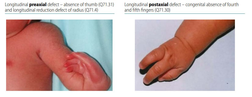
- Make sure that the preaxial side of the limb is affected – absent thumb with or without second finger, radius (upper limb); absent first with or without second toe, tibia (lower limb). Carefully distinguish from postaxial defects (see Fig. 43).
- Radiographs are very useful to characterize the bony anatomy and the missing bony segments.
- Look for signs of genetic conditions, including those that are associated with blood disorders that can be at times suspected with simple blood tests (e.g. thrombocytopenia on a complete blood count).
Checklist for high-quality reporting
| Longitudinal Preaxial Defects – Documentation Checklist |
Describe in detail, including:
|
Fig. 43. Distinguishing longitudinal preaxial defects from longitudinal postaxial defects (side-by-side comparison)

Photograph source: ECLAMC.