Common Truncus
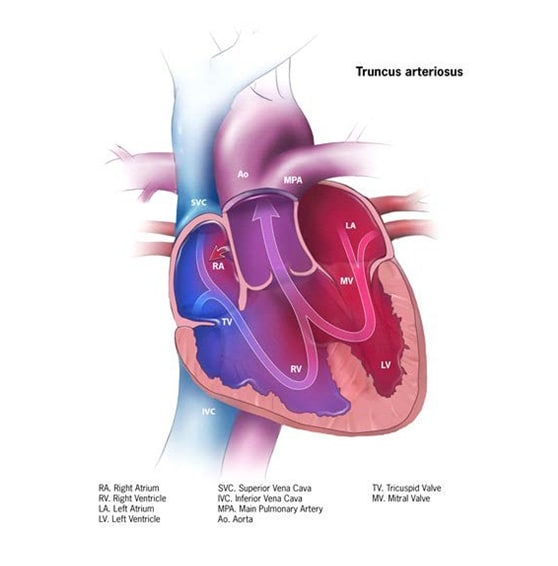
Common truncus or common arterial trunk is a structural heart defect characterized anatomically by having a single common arterial trunk, rather than a separate aorta and main pulmonary artery (see Fig. 14). This common trunk carries blood from the heart to the body, lungs, and the heart itself – that is, the common trunk gives rise to the systemic, pulmonary and coronary circulation. A ventricular septal defect is always present. Other terms for the condition are (persistent) truncus arteriosus.
Fig. 14. Common truncus


- Common truncus instead of separate aorta and main pulmonary artery.
- Ventricular septal defect, typically “high”.
- Common truncal valve.
- Variable origin of the pulmonary arteries from the common trunk.
- Frequently associated extracardiac anomalies and genetic syndromes, especially deletion 22q11.
Diagnosis
Prenatal. Common truncus can be diagnosed prenatally but might be missed or misdiagnosed, and therefore must be confirmed postnatally, typically by echocardiography.
Postnatal. The presentation after birth varies and might include a combination of heart failure (fast breathing, fast heart rate, poor feeding, and excessive sweating) and cyanosis.
Newborn screening via pulse oximetry – by non-invasively measuring blood oxygen saturation – can detect cases of common truncus if a sufficient degree of hypoxia is present at the time of screening. Echocardiography provides the specific diagnosis.
Clinical and epidemiologic notes
The clinical presentation in the newborn period might include a combination of cyanosis and heart failure.
- The clinical presentation in the newborn period might include a combination of cyanosis and heart failure.
- Pulse oximetry can identify low blood oxygen saturation and can identify cases earlier.
- Look for extracardiac anomalies (e.g. cleft palate, internal anomalies) and genetic conditions, especially deletion 22q11.
- This condition can be familial, so consider inquiring about congenital heart disease in family members.
Checklist for high-quality reporting
| Common Truncus – Documentation Checklist |
Describe in detail the clinical and echocardiographic findings:
Look for and document extracardiac birth defects: Common truncus can occur with genetic syndromes such as deletion 22q11, in which many external (e.g. cleft palate) as well as internal anomalies have been described. Report whether specialty consultation(s) have been done; in particular, whether the diagnosis was done by a paediatric cardiologist, and whether the patient was seen by a geneticist. Report genetic testing (e.g. chromosomal studies, genomic microarray, etc.) if done, and if so, report the results. |