4.2e Spina Bifida (Q05.0–Q05.9)
Spina bifida is a general term used to describe a NTD of the spine in which part of the meninges (Fig 4.7, panel a: meningocele) or spinal cord (Fig. 4.7, panel c: myelocele or myeloschisis) or both (Fig. 4.7, panel b: myelomeningocele) protrudes through an opening in the vertebral column. Spina bifida is the most common type of NTD. Hydrocephalus is a common complication, especially among children with open meningomyeloceles.
Specific types of spina bifida include:
- Meningocele: Characterized by herniation of the meninges through a spinal defect, forming a cyst filled with cerebrospinal fluid. This lesion does not contain spinal cord, but might have some nerve elements.
- Meningomyelocele (myelomeningocele): Characterized by a protrusion of the meninges and the spinal cord through an opening in the vertebral column.
- Myelocele: Characterized by a splayed vertebral column and plaque-like spinal cord without membrane or skin covering.
Fig. 4.7. Spina bifida
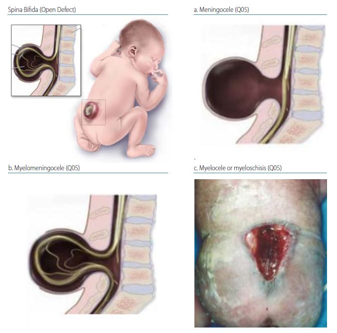
Photograph source: CDC–Beijing Medical University collaborative project.
Spina bifida lesion levels
Fig. 4.8. Cervical spina bifida
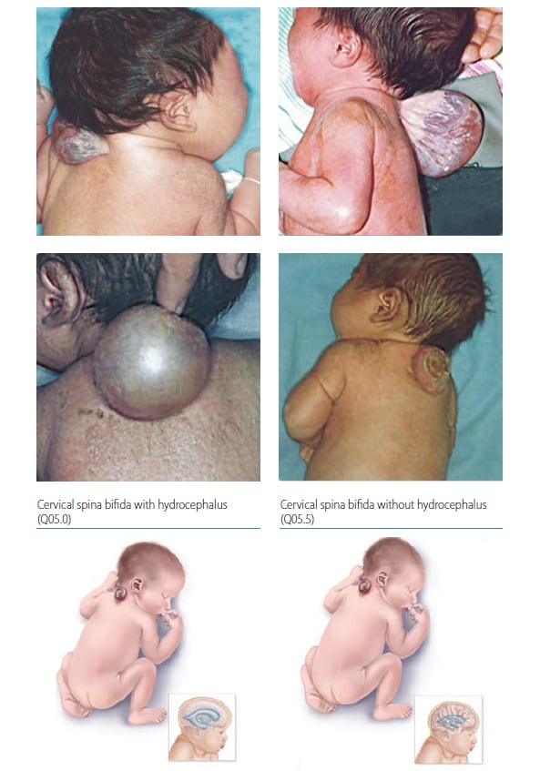
Photograph source: CDC–Beijing Medical University collaborative project.
Thoracic spina bifida
Fig. 4.9. Thoracic spina bifida
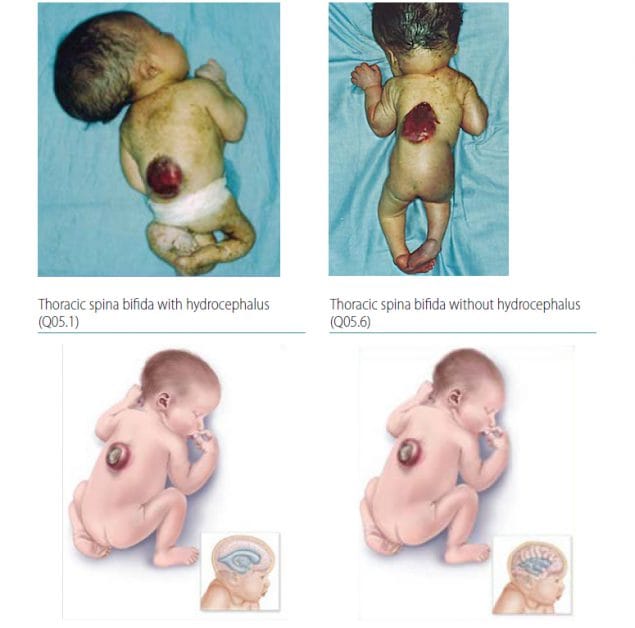
Photograph source: CDC–Beijing Medical University collaborative project.
Lumbar spina bifida
Fig. 4.10. Lumbar spina bifida
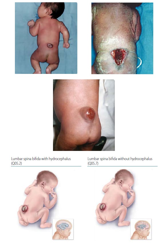
Photograph sources: CDC–Beijing Medical University collaborative project; Idalina Montes, MD, and Rafael Longo, MD, FACS, Puerto Rico.
Sacral spina bifida
Fig. 4.11. Sacral spina bifida
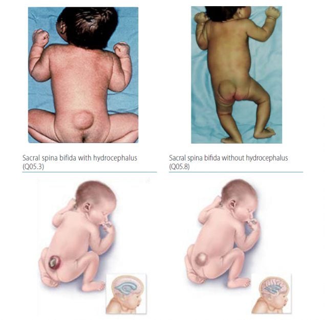
The lumbar spine is the most common location for spina bifida, followed by sacral, thoracic and cervical. Spina bifida lesions are designated as “open” if they are membrane covered and “closed” if covered by normal-appearing skin. Myelomeningocele is the most common type of spina bifida, constituting about 90% of all cases. Myelomeningoceles (which are usually open) can be clinically severe and disabling, and can cause a sequence of related findings (e.g. Chiari II malformation, hydrocephalus, hip dislocation, talipes, lower limb paralysis, and loss of sphincter control including neurogenic bladder). For infants with myelomeningocele, nerve function is intact above the lesion; therefore, lower lesions have greater preservation of neurologic function.
Infants that have meningoceles (containing only meninges and cerebral spinal fluid typically with normal structure and location of the spinal cord and nerves) experience considerably less clinical impact.
Lipomeningoceles, or lypomeningomyeloceles, are made up of a midline mass of fatty tissue (lipoma) with various amounts of tissue attached to the lower end of the spinal cord (the filum terminale). Often lipomeningo(myelo)celes are covered with a hemangioma, a patch of hair, or skin that can appear normal or have pigmentary changes. Many programmes do not classify lipomeningo(myelo)celes as an NTD. Therefore, it is critically important when developing a case definition for spina bifida to determine whether lipomeningo(myelo)celes will be included or not, and then apply the case definition consistently. If a programme chooses to include lipomeningo(myelo)celes, the most common ICD-10 Royal College of Paediatrics and Child Health (RCPCH) code used is Q05.4 – “Spina bifida unspecified”. Other terminal defects – such as myelocystocele (which might present as a sacral mass), or occult spinal dysraphisms such as split spinal cord malformation, tethered cord syndrome, or spinal lipomas, among others – are also not classified as NTDs and are coded as Q06.8 – “Other specified congenital malformations of spinal cord”. Spina bifida occulta is a defect in the posterior vertebral arches without neural involvement, most commonly at the lumbosacral junction, and is also not classified as an NTD. The RCPCH code for this defect is Q76.0 –“Spina bifidaocculta”.
Relevant ICD-10 codes
Q05.0 Cervical spina bifida with hydrocephalus
Q05.1 Thoracic spina bifida with hydrocephalus
Q05.2 Lumbar spina bifida with hydrocephalus (includes lumbosacral spina bifida with hydrocephalus)
Q05.3 Sacral spina bifida with hydrocephalus
Q05.4 Unspecified spina bifida with hydrocephalus
Q05.5 Cervical spina bifida without hydrocephalus
Q05.6 Thoracic spina bifida without hydrocephalus
Q05.7 Lumbar spina bifida without hydrocephalus (includes lumbosacral spina bifida without hydrocephalus)
Q05.8 Sacral spina bifida with hydrocephalus
Q05.9 Spina bifida, unspecified
Diagnosis
Prenatal. Spina bifida might be diagnosed prenatally using ultrasound. However, if the entire spine is difficult to image, distinguishing whether the lesion is open or closed is challenging. Results of maternal serum screening for alpha fetoprotein (AFP), if available, might help to determine if the lesion is open or closed, as AFP seeps out of the open lesion into the amniotic fluid and subsequently into the mother’s blood. Spina bifida is sometimes confused with sacrococcygeal teratoma, isolated scoliosis/kyphosis or amniotic band syndrome. Therefore, a prenatal diagnosis of spina bifida should be confirmed postnatally. When this is not possible (e.g. termination of pregnancy or unexamined fetal death), the programme should have criteria in place to determine whether to accept or not accept a case based solely on prenatal data.
Postnatal. The newborn examination usually confirms the diagnosis. Neurologic impairment will vary by spina bifida type, level of lesion and severity. Imaging (when available) can provide additional information to characterize the location, extent and content of the lesion, as well as the presence or absence of frequently cooccurring brain findings (e.g. hydrocephalus, Chiari II malformation).
Clinical and epidemiologic notes
Distinguishing spina bifida from the other abnormalities of the spine is important because these conditions have different causes and associated anomalies. With careful examination, the diagnosis of spina bifida is straightforward, but imaging (when available) is very helpful. The extent of the spinal dysraphism has been shown to extend above the visible lesion and the highest level per x-ray should be coded. Conditions that might be misdiagnosed as spina bifida either prenatally or postnatally include sacrococcygeal teratoma, isolated scoliosis/kyphosis and amniotic band syndrome.
Spina bifida is often an isolated, non-syndromic anomaly. For this reason, it is crucial to report all findings and obtain good clinical photographs for the expert reviewer. However, spina bifida can occur with genetic syndromes (e.g. trisomy 18) and is occasionally a manifestation of single-gene disorders such as Waardenburg syndrome.
Non-genetic risk factors include pregestational diabetes, obesity, seizure medications (i.e. valproic acid, carbamazepine), hyperthermia (e.g. fever) in early pregnancy, and folic acid insufficiency/deficiency. Adequate periconceptional use of folic acid (as a supplement or through fortification) can prevent most cases of spina bifida.
The birth prevalence of spina bifida varies widely, between 0.6 to 38.9 per 10 000 births. Prevalence is higher among women of Hispanic ancestry compared to non-Hispanic white and non-Hispanic black women. Lower-income countries and countries without mandated folic acid fortification of staple foods have a higher prevalence of spina bifida.
Inclusions
Q05.0 Cervical spina bifida with hydrocephalus
Q05.1 Thoracic spina bifida with hydrocephalus
Q05.2 Lumbar spina bifida with hydrocephalus (includes lumbosacral spina bifida with hydrocephalus)
Q05.3 Sacral spina bifida with hydrocephalus
Q05.4 Unspecified spina bifida with hydrocephalus
Q05.5 Cervical spina bifida without hydrocephalus
Q05.6 Thoracic spina bifida without hydrocephalus
Q05.7 Lumbar spina bifida without hydrocephalus (includes lumbosacral spina bifida without hydrocephalus)
Q05.8 Sacral spina bifida with hydrocephalus
Q05.9 Spina bifida, unspecified
Exclusions
D48 Neoplasm of uncertain behavior (sacrococcygeal teratoma)
Q06.8 Other specified congenital malformations of spinal cord
Q07 Other congenital malformations of nervous system
Q76.0 Spina bifida occulta
Checklist for high-quality reporting
| Spina Bifida – Documentation Checklist |
Describe defect in detail:
Report whether autopsy (pathology) findings are available and if so, report the results. |
Suggested data quality indicators
| Category | Suggested Practices and Quality indicators |
| Description and documentation |
|
| Coding |
|
| Clinical classification |
|
| Prevalence |
|