Congen Anom of the Dig Sys: Oesophageal Atresia/Tracheo-Oesophageal Fistula
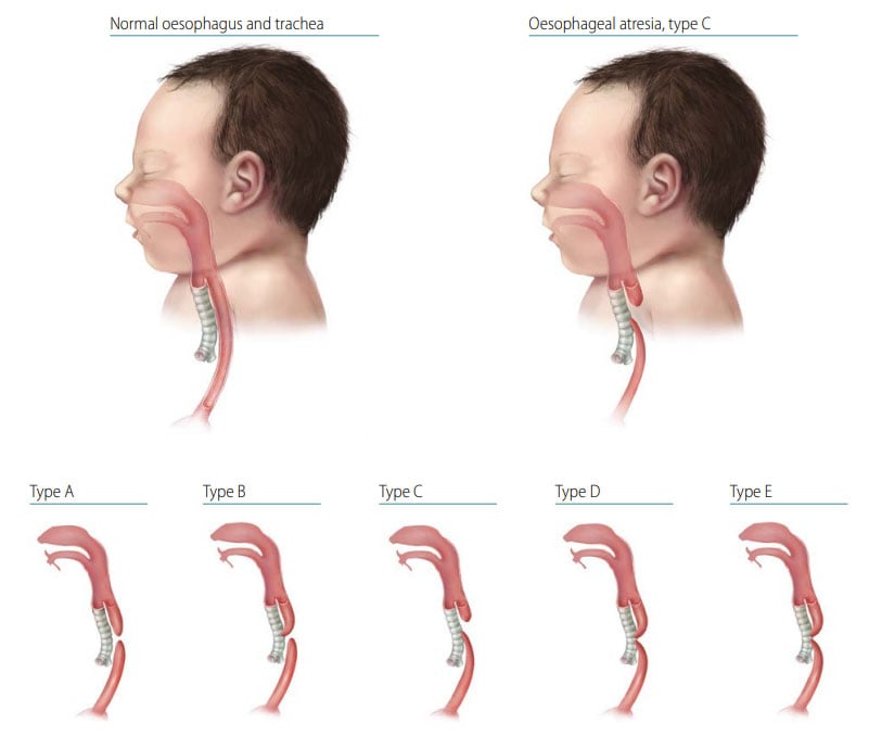
Oesophageal (esophageal) atresia is a congenital malformation characterized by the oesophagus ending in a blind pouch that does not connect to the stomach. Tracheo-oesophageal fistula (TEF or TOF) consists of a communication between the oesophagus and the trachea that is not normally present. Although it might occur alone, TEF is commonly associated with oesophageal atresia.
Fig. 29. Oesophageal atresia/tracheo-oesophageal fistula

-
- The anatomical types of oesophageal atresia are:
- Type A: Oesophageal atresia without TEF.
- Type B: Oesophageal atresia with proximal TEF.
- Type C: Oesophageal atresia with distal TEF.
- Type D: Oesophageal atresia with proximal and distal TEF.
- Type E: TEF without oesophageal atresia.
Types A–D are typically recognized oesophageal atresia, with type C being by far the most common.
- Signs of oesophageal atresia at birth include vomiting immediately after feeding, excessive drooling or mucus, and, if TEF is present, respiratory distress.
- The anatomical types of oesophageal atresia are:
Diagnosis
Prenatal. Oesophageal atresia is difficult to diagnose prenatally. Always confirm a prenatal diagnosis.
Postnatal. Oesophageal atresia can be diagnosed radiographically when a feeding tube cannot pass from the pharynx into the stomach and, if there is no TEF, by absence of air in the stomach. Because of the variability in symptoms, the diagnosis of TEF without oesophageal atresia may be delayed for weeks, months, or even years.
Clinical and epidemiologic notes
- Other unrelated birth defects, particularly cardiac, anorectal, skeletal/vertebral and urogenital.
- Anomaly complexes (e.g. OAVS [oculo-auriculo-vertebral spectrum], VATER or VACTERL association [vertebral, anus, cardiac, trachea, oesophagus, renal, limb]).
- Genetic syndromes (e.g. trisomies 18 and 21, CHARGE [coloboma, heart defects, choanal atresia, growth retardation, genital abnormalities, ear abnormalities] syndrome, Feingold syndrome).
- Always look for additional internal anomalies and syndromes.
- Note the type of oesophageal atresia and if TEF occurs.
Checklist for high-quality reporting
| Oesophageal Atresia with or without TEF – Documentation Checklist |
Describe in detail:
Describe evaluations to rule out additional malformations/associations/syndromes:
Take and report photographs: Show clearly oesophageal atresia and, if present, TEF type; can be crucial for review. Report whether autopsy (pathology) findings are available and if so, report the results. |