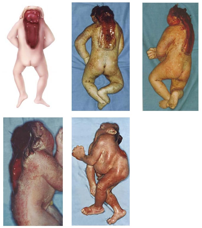
Craniorachischisis
Craniorachischisis is characterized by the combination of anencephaly (absence of the brain and cranial vault, without skin covering) with a contiguous bony defect of the cervical spine (also without meninges covering the neural tissue – rachischisis).
Fig. 3. Craniorachischisis

Photograph source: CDC–Beijing Medical University collaborative project.
- Head – anencephaly (absence of the brain and cranial vault).
- Covering – no skin covering residual brain tissue, spinal cord tissue, or cranial vault (calvarium).
- Spine – open (rachischisis) might be limited to the cervical spine, but the open defect can extend to the thoracic spine or even lumbar or sacral spine (craniorachischisis totalis).
Diagnosis
Prenatal. Craniorachischisis is readily diagnosed using ultrasound but can be confused with other defects involving the brain – anencephaly, acrania and amniotic band syndrome. Use programme rules (SOPs) to decide whether to accept or not accept prenatal diagnoses without postnatal confirmation (e.g. in cases of termination of pregnancy or unexamined fetal death).
Postnatal. Careful examination of the fetus or newborn can confirm the diagnosis of craniorachischisis and distinguish it from the other rare anomalies that involve the brain, cranium and spine.
Clinical and epidemiologic notes
- Eyes are normally formed; bulging is a result of absence of the frontal portion of the cranial vault.
- Neck may appear to be shortened and is sometimes retroflexed.
- Cerebellum and brain stem are intact.
- Craniorachischisis is always an open lesion, with the anencephaly always being contiguous with the spinal lesion.
- Craniorachischisis might co-occur with other anomalies: Cleft lip and palate, omphalocele, limb defects, or cyclopia.
- Check for chromosomal anomaly: Case reports of trisomy 18 with craniorachischisis and omphalocele have been reported.
- Craniorachischisis might be confused with:
- Amniotic band or limb-body wall spectrum, which have other findings (facial schisis, limb and ventral wall anomalies, bands) and allow the differentiation from typical craniorachischisis.
- Iniencephaly if spinal retroflexion is present.
Checklist for high-quality reporting
| Craniorachischisis – Documentation Checklist |
Describe in detail:
Take and report photographs: Show clearly the missing cranium; can be crucial for review. Describe evaluations to find or rule out related and associated anomalies:
Report whether autopsy (pathology) findings are available and if so, report the results. |