Orofacial Clefts: Cleft Palate
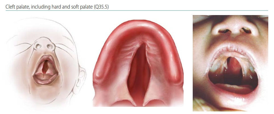
Cleft palate (also called palatoschisis) is characterized by a fissure (clefting) in the secondary palate (posterior to the incisive foramen) and can involve the soft palate only (the most posterior part of the palate), or both the hard palate and the soft palate. The cleft can be narrow (V-shaped), or wider (U-shaped) (see Fig. 21). The lip is intact.
Fig. 21. Cleft palate

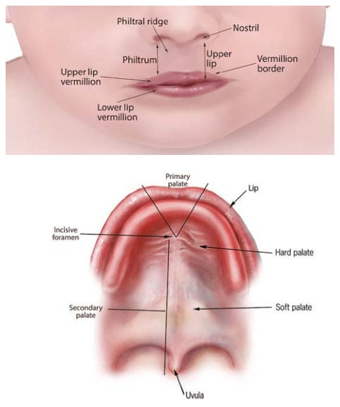
Fig. 22. Anatomy of the lip and palate

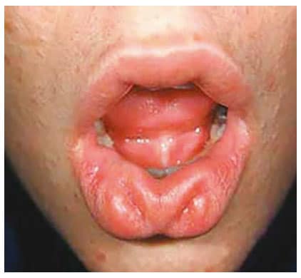
Fig. 23. Lip pits

Diagnosis
Prenatal. Cleft palate alone can be suspected prenatally but can easily be missed or misdiagnosed. Cases identified or suspected prenatally should be confirmed postnatally before inclusion in a surveillance programme.
Postnatal. Cleft palate can be missed at the external newborn examination if the palate is not systematically and carefully examined. This requires visualization of the entire length of the palate.
Clinical and epidemiologic notes
In cleft palate, a complete evaluation and physical examination is crucial as it is more commonly associated with additional anomalies and syndromes compared to other types of clefts (e.g. cleft lip).
- Check for lip pits in the lower lip (Fig. 23), in the child and in the parents – it is a sign of a genetic condition (van der Woude syndrome) with high recurrence risk (a parent may have the pits but not the cleft).
- Check for additional anomalies, especially of the heart (e.g. in deletion 22q11 syndrome) and eye (e.g. Stickler syndrome).
- Check for components of the Pierre Robin sequence, including microretrognathia (small recessed jaw), glossoptosis (posterior displacement of the tongue) and respiratory obstruction.
Checklist for high-quality reporting
| Cleft Palate – Documentation Checklist |
Describe in detail, including:
Describe procedures to assess further additional malformations and if present, describe. Take and report photographs: Very useful; can be crucial for review. Report whether specialty consultation(s) were done and if so, report the results. Plastic surgery and genetics consultation reports are useful. |
Key visuals:
Anatomy of the lip and palate. Note that in bilateral cleft lip, a median remnant of the philtrum is still present. Note also that the clefting can extend to the gum or alveolus but does not extend beyond the incisive foramen (thus involving only the primary palate) (see Fig. 22).
Lip pits. Lip “pits” are indentations, often with raised borders, on the lower lip (see Fig. 23). They are a sign of specific syndromes (most commonly van der Woude syndrome).