4.5 Congenital Malformations and Deformations of the Musculoskeletal System
On this Page
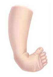
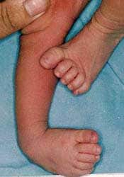
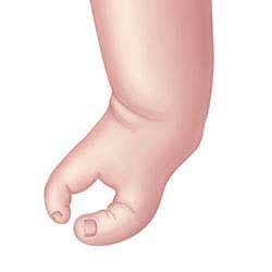
Talipes equinovarus (Q66.0, Q66.1, Q66.4, Q66.8)
The term “clubfoot” is sometimes used to describe several kinds of ankle or foot defects present at birth. However, orthopaedic specialists use it as a synonym for talipes equinovarus (see Fig. 4.12). The condition, which has a wide spectrum of severity, is characterized by adduction of the forefoot and midfoot, adduction of the heel or hind foot, and a fixed plantar flexion (equinus position) of the ankle (29). In other words, the foot points downward and inward and is rotated outward axially. Other defects of the foot and ankle include talipes calcaneovalgus (in which the ankle joint is dorsiflexed and the forefoot deviated outwards) and talipes calcaneovarus (in which the ankle joint is dorsiflexed and the forefoot deviated inwards).
| Relevant ICD-10 codes and Royal College of Paediatrics and Child Health (RCPCH) uses: | |
| Q66 | Congenital deformities of feet (avoid using this general code if more specific information is available) |
| Q66.0 | Talipes equinovarus |
| Q66.1 | Talipes calcaneovarus |
| Q66.4 | Talipes calcaneovalgus |
| Q66.8 | Other congenital deformities of feet |
| Clubfoot NOS (not otherwise specified) | |
| Exclusions | |
| Clubfoot, positional (no associated ICD-10 codes) | |
| Clubfoot associated with neuromuscular diagnoses or syndromes, such as arthrogryposis multiplex congenital, congenital myotonic dystrophy, and diastrophic dysplasia (no associated ICD-10 codes) |
Reduction defects of upper and lower limbs (longitudinal, transverse and intercalary)
Limb deficiencies make up a large number of defects in limb development, characterized by the total or partial absence or different degrees of hypoplasia of the skeletal structures of the limbs. They are classified into three large groups: longitudinal, transverse and intercalary limb deficiencies. Some cases will have multiple limb defects, and therefore will be classified in more than one of these groups.
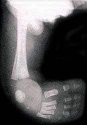
Longitudinal limb deficiencies (Q71.4, Q71.5, Q71.6, Q72.4, Q72.5, Q72.6, Q72.7)
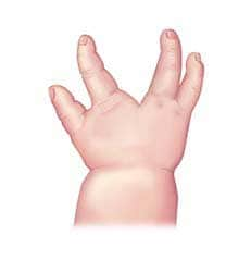
Longitudinal limb deficiencies (see Fig. 4.13–4.18) refer to the partial absence of a limb extending parallel to the long axis of the limb. They typically involve specific components of the limbs: preaxial (first ray: thumb or radius in the arm(s), or both, or first toe or tibia in the leg(s), or both); postaxial (fifth ray: fifth finger or ulna in the arm(s), or both, fifth toe or fibula in the leg(s), or both); or central components (typically, third or fourth rays in the hand(s) (also called split hand or lobster-claw hand) or foot (also called split foot), or both.
Fig. 4.13. Radial aplasia
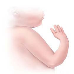
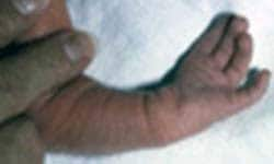
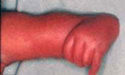
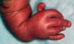
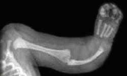
Fig. 4.14. Ulnar aplasia/hypoplasia
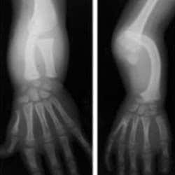
Fig. 4.15. Femoral and fibular aplasia
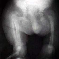
Fig. 4.16. Tibial aplasia
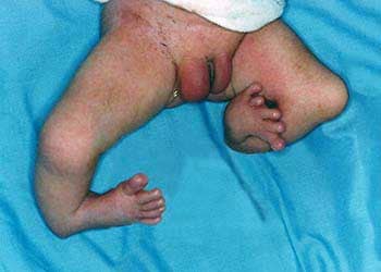
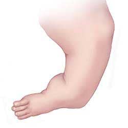
Fig. 4.17. Split hand
Transverse limb deficiencies (Q71.0, Q71.2, Q71.3, Q72.0, Q72.2, Q72.3)
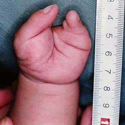
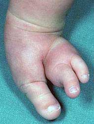
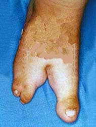
Transverse limb deficiencies (see Fig. 4.19–4.29) refer to the complete or partial absence of distal structures of a limb in a transverse plane at the point where the deficiency begins, with proximal structures being essentially intact. Transverse limb deficiencies also are known as congenital amputations.
Fig. 4.19. Congenital absence of forearm and hand
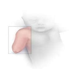
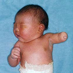
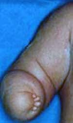
Fig. 4.20. Congenitalabsence of both forearm and hand
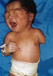
Fig. 4.21. Congenital absence of lower leg and foot
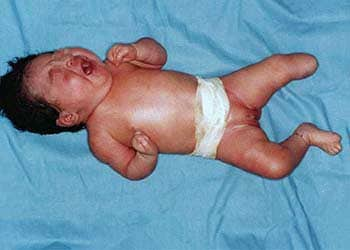
Fig. 4.22. Aphalangia of the hand. Partial absence of the phalanges
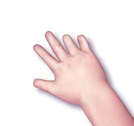
Fig. 4.23. Aphalangia of the feet. Partial absence of the phalanges
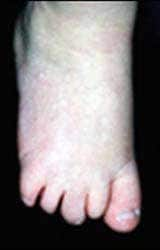
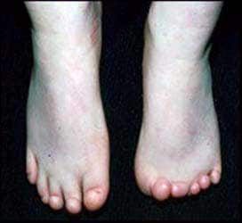
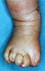
Fig. 4.24. Adactyly of the hand
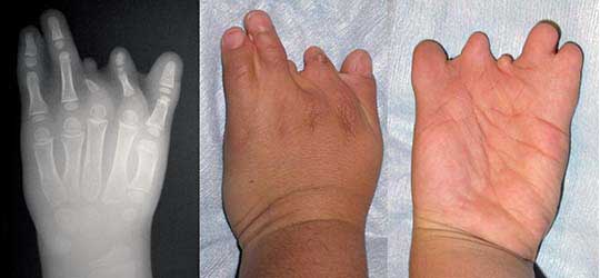
Fig. 4.25. Adactyly of the feet
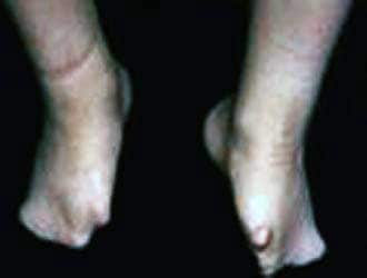
Fig. 4.26. Oligodactyly of the hand
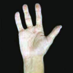
Fig. 4.27. Oligodactyly of the foot (absent hallux)
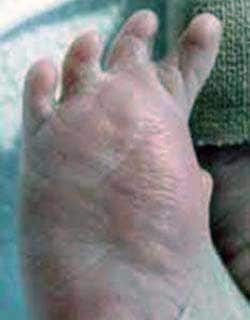
Fig. 4.28. Amelia upper limb
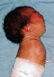
Fig. 4.29. Amelia of the lower limb
Intercalary limb deficiencies (Q71.1 and Q72.1)
Intercalary limb deficiencies refer to the complete or partial absence of the proximal or middle segment(s) of a limb, with all or part of the distal segment present (see Fig. 4.30-4.31).
Fig. 4.30. Reduction defects of upper arm and forearm with hand present
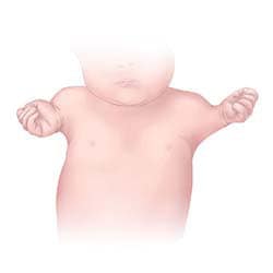
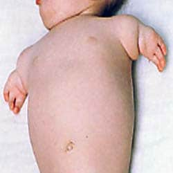
Fig. 4.31. Reduction defects of thigh and lower leg with foot present
| Relevant ICD-10 codes and RCPCH uses: | |
| Reduction defects of upper limbs | |
| Q71 | Reduction defects of upper limb (avoid using this general code if more specific information is available) |
| Q71.0 | Congenital complete absence of upper limb(s) |
| Amelia of upper limb | |
| Congenital absence of upper arm and forearm with hand present | |
| Q71.1 | Phocomelia of upper limb |
| Congenital absence of both forearm and hand | |
| Q71.3 | Congenital absence of hand and finger(s) |
| Q71.30 | Congenital absence of finger(s) (Remainder of hand intact) |
| Aphalangia: absent phalanx (an individual bone in a finger) or phalanges | |
| Adactyly: absence of fingers (generally refers to all fingers on a hand, although soft tissue nubbins without bones can be present | |
| Oligodactyly: fewer than 10 complete fingers | |
| Q71.31 | Absence or hypoplasia of thumb (Other digits intact) |
| Q71.4 | Longitudinal reduction defect of radius |
| Radial aplasia/hypoplasia | |
| Clubhand (congenital) | |
| Radial clubhand | |
| Q71.5 | Longitudinal reduction defect of ulna |
| Ulnar aplasia/hypoplasia | |
| Q71.6 | Lobster-claw hand |
| Split hand | |
| Congenital cleft hand | |
| Q71.8 | Other reduction defects of upper limb(s) |
| Congenital shortening of upper limb(s) | |
| Q71.9 | Reduction defect of upper limb, unspecified |
| Reduction defects of lower limbs | |
| Q72 | Reduction defects of lower limb (avoid using this general code if more specific information is available) |
| Q72.0 | Congenital complete absence of lower limb(s) |
| Amelia of lower limb | |
| Q72.1 | Congenital absence of thigh and lower leg with foot present |
| Phocomelia of lower limb | |
| Q72.2 | Congenital absence of both lower leg and foot |
| Q72.3 | Congenital absence of foot and toe(s) |
| Q72.30 | Congenital absence or hypoplasia of toe (s) with remainder of foot intact |
| Aphalangia: absent phalanx (an individual bone in a toe) or phalanges | |
| Adactyly: absence of toes (generally refers to all toes on a foot, although soft tissue nubbins without bones can be present) | |
| Oligodactyly: fewer than 10 complete toes | |
| Q72.31 | Absence or hypoplasia of first toe with other digits present |
| Q72.4 | Longitudinal reduction defect of femur (commonly referred to as femoral aplasia/hypoplasia) |
| Proximal femoral focal deficiency | |
| Q72.5 | Longitudinal reduction defect of tibia |
| Tibial aplasia/hypoplasia | |
| Q72.6 | Longitudinal reduction defect of fibula |
| Fibular aplasia/hypoplasia | |
| Q72.7 | Split foot |
| Q72.8 | Other reduction defects of lower limb(s) |
| Congenital shortening of lower limb(s) | |
| Q72.9 | Reduction defect of lower limb, unspecified |
| Reduction defect of unspecified limb | |
| Q73 | Reduction defects of unspecified limb (avoid using this general code if more specific information is available) |
| Q73.0 | Congenital absence of unspecified limb(s) |
| Amelia NOS | |
| Q73.1 | Phocomelia, unspecified limb(s) |
| Phocomelia NOS | |
| Q73.8 | Other reduction defects of unspecified limb(s) |
| Longitudinal reduction deformity of unspecified limb(s) | |
| Ectromelia NOS | |
| Hemimelia NOS | |
| Reduction defect NOS | |
| Q87.2 | Sirenomelia |
| Exclusions | |
| Q77.0–Q77.9, Q78.0–Q78.9 | Generalized limb shortening including skeletal dysplasias (osteochondrodysplasias) |
| Q79.80 | Cases with known or probable amniotic band/constriction band presencea |
| Q84.6 | Nail hypoplasia |
| Q89.80 | Lower extremity deficiencies with caudal dysgenesis |
| Q73.80 | Absent digits, unspecified |
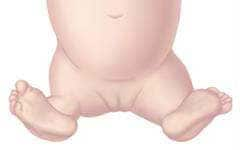
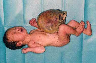
Exomphalos/omphalocele (Q79.2)
Omphalocele (see Fig. 4.32) is a birth defect of the anterior abdominal wall, in which the herniated intestines and abdominal organs are usually covered by a membrane consisting of the peritoneum and amnion. In contrast to gastroschisis, in which the abdominal defect is lateral to the umbilicus, in omphalocele the abdominal contents are herniated through an enlarged umbilical ring and the umbilical cord is inserted in the distal part of the membrane covering the defect.
| Relevant ICD-10 codes and RCPCH uses: | |
| Q79 | Congenital malformations of the musculoskeletal system, not elsewhere classified |
| (avoid using this general code if more specific information is available) | |
| Q79.2 | Exomphalos/omphalocele |
| Exclusions | |
| Q79.3 | Gastroschisis |
| Q79.8 | Umbilical hernia |
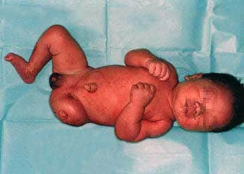
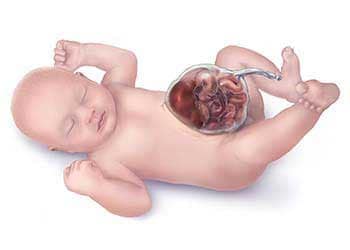
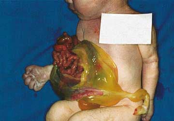
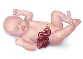
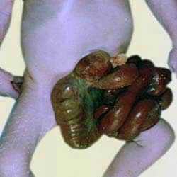
Gastroschisis (Q79.3)
Gastroschisis is also a birth defect of the anterior abdominal wall, accompanied by herniation of the small intestine and part of the large intestine, and occasionally other abdominal organs, into the amniotic cavity (see Fig. 4.33). Importantly, the herniated organs lack a protective membrane. The extruded abdominal contents can be matted and covered by a thick fibrous material, but this membrane does not resemble skin. Gastroschisis occurs lateral to the umbilicus (generally to the right).
Gastroschisis and omphalocele can be confused with one another when the membrane covering the omphalocele has ruptured. However, careful examination demonstrating the position of the abdominal opening lateral to the umbilical cord insertion helps confirm the diagnosis of gastroschisis.
Fig. 4.33. Gastroschisis
| Relevant ICD-10 codes and RCPCH uses: | |
| Q79 | Congenital malformations of the musculoskeletal system, not elsewhere classified |
| (avoid using this general code if more specific information is available) | |
| Q79.3 | Gastroschisis |
| Exclusions | |
| Q79.2 | Exomphalos/omphalocele |
| Q89.81 | Limb–body wall complex |
- Photograph and X-ray source: courtesy of Idalina Montes, MD and Rafael Longo, MD, FACS, Puerto Rico.
- Photographs source: courtesy of CDC-Beijing Medical University collaborative project.
- Photograph source and X-Ray source: courtesy of Jaime Frías, MD, USA.
- Photograph source: courtesy of Estudio Colaborativo Latino Americano de Malformaciones Congénitas (ECLAMC).
- Photograph source: courtesy of John Wiley and Sons ©2009. Biesecker LG et al. Am. J. Med. Genet. A. 2009;149A:93–127.
- Photographs and X-ray source: courtesy of Dr E Gene Deune, MD; Associate Professor, Johns Hopkins Department of Orthopedic Surgery, Division of Hand Surgery, Baltimore, MD, USA.
- X-ray source: courtesy of John Wiley and Sons ©2011. Umaña LA et al. Am. J. Med. Genet. A. 2011;155A:3071–4.
- X-ray source: courtesy of John Wiley and Sons ©1997. Kumar D et al. Am. J. Med. Genet. A. 1997;70A:107–13.