Case #490 – April, 2019
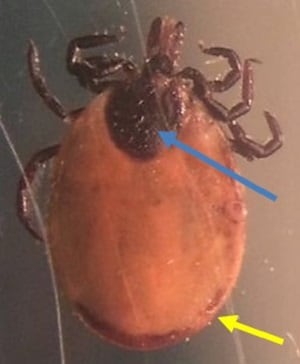
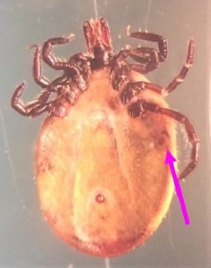
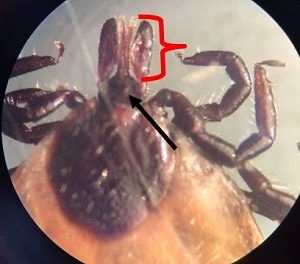
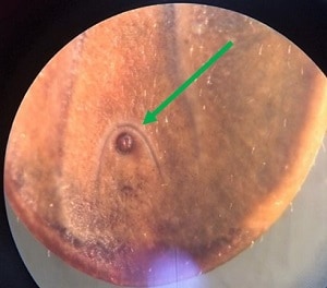
A 55-year old man from Massachusetts removed a tick attached to his forearm after returning from a weekend camping trip. He then took it to his county health clinic from where it was subsequently sent to the State Public Health laboratory for identification. Figure A shows a dorsal view of the tick, while Figure B shows the ventral side. Figure C and D shows a close-up of the mouthparts while Figure E is a close-up of the lower ventral side. What is your identification? Based on what criteria? What is the public health importance, if any, of this genus in North America?
These images were kindly provided by Amanda Castonguay from Brigham and Women’s Hospital, Boston, MA

Figure A
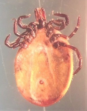
Figure B
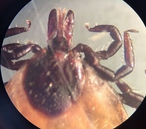
Figure C
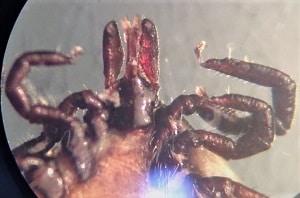
Figure D
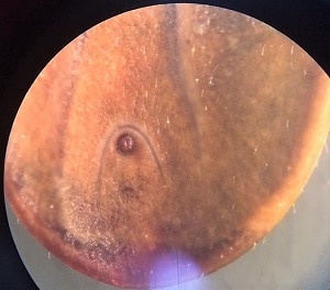
Figure E
Images presented in the dpdx case studies are from specimens submitted for diagnosis or archiving. On rare occasions, clinical histories given may be partly fictitious.
DPDx is an educational resource designed for health professionals and laboratory scientists. For an overview including prevention, control, and treatment visit www.cdc.gov/parasites/.
