
Case #479 – November, 2018
A 29-year-old female with a nodule on her left back was examined at a university hospital in Belgium. The nodule was excised and sent to pathology. Figures A – C show what was observed on hematoxylin- and eosin-stained sections of the nodule. What is your diagnosis? Based on what criteria?
(This case and images were kindly provided by the Antwerp University Hospital (UZA), Department of Pathological Anatomy, Antwerp, Belgium)

Figure A

Figure B

Figure C
This was a case of dirofilariasis caused by Dirofilaria repens. In addition to the geographical region where the infection was acquired (Old World), the following morphologic features were also shown in the Figures:
- A thick cuticle with cuticular ridges present in some areas
- Short coelomyarian muscle cells
- An internal lateral ridge (black arrows, Figure B)
- Lack of external alae
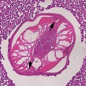
Figure B
More on dirofilariasis: https://www.cdc.gov/dpdx/dirofilariasis/index.html
Images presented in the dpdx case studies are from specimens submitted for diagnosis or archiving. On rare occasions, clinical histories given may be partly fictitious.
DPDx is an educational resource designed for health professionals and laboratory scientists. For an overview including prevention, control, and treatment visit www.cdc.gov/parasites/.