
Case #453 – October, 2017
A 38-year-old asplenic man residing in California was hospitalized with a 104º F fever, hematuria, myalgia, fatigue and thrombocytopenia. The patient had recently traveled to Arizona, Kansas and Oklahoma. A blood specimen collected in EDTA was sent to the hematology lab for routine work-up. Images were captured and sent to the DPDx Team for diagnostic assistance. Figures A – C shows what was observed by the technician in a thin smear for differential WBC count. What is your diagnosis? Based on what morphologic features?
This case and images were kindly provided by the California Public Health Laboratory, Richmond, California.

Figure A

Figure B

Figure C
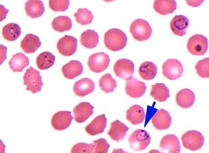
Figure A
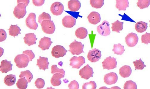
Figure B
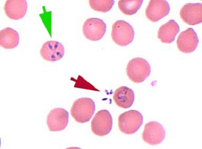
Figure C
This was a case of babesiosis caused by Babesia sp. Morphologic features included:
- Absence of malarial pigment found in Plasmodium species.
- Pleomorphic ring forms in normal-sized red blood cells Figures A and B (blue arrows).
- Characteristic tetrad (Maltese cross) forms Figures B and C (green arrows).
- Extracellular form Figure C (red arrow).
- Presence of only rings and ring-like forms.
In general, identification of Babesia to the species or strain level is not possible by morphology, and requires molecular analysis of a blood specimen.
More on: Babesiosis
Images presented in the dpdx case studies are from specimens submitted for diagnosis or archiving. On rare occasions, clinical histories given may be partly fictitious.
DPDx is an educational resource designed for health professionals and laboratory scientists. For an overview including prevention, control, and treatment visit www.cdc.gov/parasites/.