
Case #188 – September, 2006
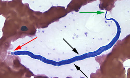
A hospital submitted blood films to CDC’s reference laboratory for identification of the microfilariae that were seen on the films. The object in Figure A was seen on a Wright-Giemsa stained blood film and measured approximately 180 µm. The object in B was seen on a Giemsa stained blood film and measured approximately 240-250 µm. Both A and B were taken at 500× magnification. What is your diagnosis? Based on what criteria?

Figure A

Figure B
Images presented in the DPDx case studies are from specimens submitted for diagnosis or archiving. On rare occasions, clinical histories given may be partly fictitious.
DPDx is an educational resource designed for health professionals and laboratory scientists. For an overview including prevention, control, and treatment visit www.cdc.gov/parasites/.