Case #494 – June, 2019
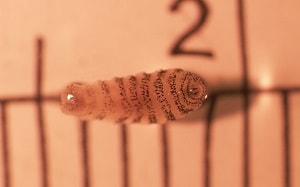
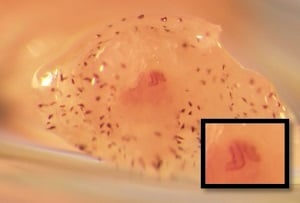
A 30-year-old stable worker from New Hampshire sought medical attention at a local health center for a painful bump with cellulitis on the side of her neck. The bump failed to go away with antibiotics, and when the wound was drained, the 3.5 mm long object pictured below was produced. Figure A shows the entire parasite, and Figure B shows a view straight down onto the posterior end of the organism. Of note: for Figure B, the terminal segment has withdrawn inside the preceding segment, making visualization of the diagnostic features very difficult. Only one of the paired spiracular plates is visible (inset).
What is your diagnosis? Based on what criteria?

Figure A

Figure B
Images presented in the dpdx case studies are from specimens submitted for diagnosis or archiving. On rare occasions, clinical histories given may be partly fictitious.
DPDx is an educational resource designed for health professionals and laboratory scientists. For an overview including prevention, control, and treatment visit www.cdc.gov/parasites/.