
Case #471 – July, 2018
A 79 year-old man was diagnosed with encephalitis at a local hospital in Arizona. A Computed Tomography (CT) scan showed multiple brain lesions. A biopsy specimen from the occipital lobe was collected and sent to Pathology for routine histological workup. Images of structures shown in Figures A, B and C were observed by the pathologist on hematoxylin-and-eosin (H&E) stained sections and sent to DPDx for diagnostic assistance. The objects of interest shown in the Figures measured 10 – 25 µm in diameter. What is your diagnosis? Based on what criteria?
This case and the images were kindly contributed by the Health-Banner Desert Hospital, Mesa AZ

Figure A

Figure B

Figure C
This was a case of granulomatous amebic encephalitis (GAE) caused by Balamuthia mandrillaris. Diagnostic features were:
- the pathology of the infection, manifesting as a rapid granulomatous encephalitis.
- the presence of thick-walled cysts (Figure A, blue arrow) that are hexagonal to polygonal (Figure C, green arrows).
Balamuthia is one of three genera of typically free-living amebae implicated in amebic encephalitis, the other two being Naegleria and Acanthamoeba. Amebae enter the body through nasal passages or broken skin and can invade the central nervous system by hematogenous dissemination. Identification to the genus level is sometimes difficult based on morphology of the amebae alone. This case was confirmed by real-time PCR using Balamuthia-specific primers.
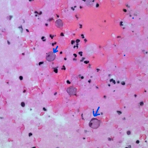
Figure A

Figure C
For more information on Balamuthia visit: https://www.cdc.gov/dpdx/freelivingamebic/index.html
Images presented in the dpdx case studies are from specimens submitted for diagnosis or archiving. On rare occasions, clinical histories given may be partly fictitious.
DPDx is an educational resource designed for health professionals and laboratory scientists. For an overview including prevention, control, and treatment visit www.cdc.gov/parasites/.