Case #466 – April, 2018
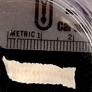
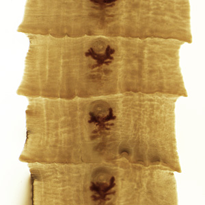
A 6-year-old girl, who originally lived in Japan but now resides in the United States, was seen by a health care provider for abdominal pain and diarrhea. A stool specimen was collected and the object shown in Figure A was observed in the specimen. It was sent to the CDC DPDx Team for identification. The object was cleared using lacto-phenol and Figure B shows what was observed using a dissecting microscope. What is your diagnosis? Based on what criteria? Would you suggest any other test(s)?

Figure A

Figure B
Images presented in the dpdx case studies are from specimens submitted for diagnosis or archiving. On rare occasions, clinical histories given may be partly fictitious.
DPDx is an educational resource designed for health professionals and laboratory scientists. For an overview including prevention, control, and treatment visit www.cdc.gov/parasites/.