
Case #465 – April, 2018
A 74-year-old man, with a prior history of urothelial carcinoma, high grade pT1 (invasive, into lamina propria), was seen at a medical facility for follow-up care. Travel history for the patient was not available. A urinary cytology specimen was examined by a pathologist who observed structures thought to be parasitic in nature. Digital images were captured and sent to the CDC DPDx Team for diagnostic assistance. Figures A and B represent the structures that were observed. What is your diagnosis? Based on what morphologic features.
This case and images were kindly provided by the Hospital Professor Doutor Fernando Fonseca, Amadora, Portugal.

Figure A

Figure B
The objects were variations of crystals of uric acid so the diagnosis was No Parasites Found. Morphologic features shown which excluded a diagnosis of Schistosoma haematobium included:
- size of the objects (although not given) being considerable smaller than eggs of Schistosoma haematobium. Observation of the epithelial cells present allows for an estimation of size.
- absence of a miracidium.
- lack of a single terminal spine; some objects appeared to have a point on both ends.
The submitter also employed polarized microscopy, which showed that the crystals were highly birefringent (double refractive) which also helps to distinguish crystals of uric acid from those of cysteine (Figures A and B).
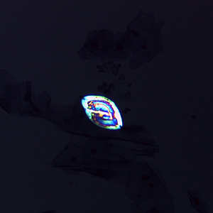
Figure A
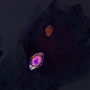
Figure B
Images presented in the dpdx case studies are from specimens submitted for diagnosis or archiving. On rare occasions, clinical histories given may be partly fictitious.
DPDx is an educational resource designed for health professionals and laboratory scientists. For an overview including prevention, control, and treatment visit www.cdc.gov/parasites/.