
Case #462 – February, 2018
A 17 year-old male patient presented with cellulitis and a mass in the eye over a 3 month period. It was situated at the limbus near the lateral rectus muscle. Surgical exploration revealed a sub-conjunctival cystic mass about 1 cm in diameter. The mass was subsequently excised and sent to the pathologist for examination. The attached images were taken from hematoxylin and eosin (H&E) stained sections. What is your diagnosis? Based on what criteria?
This case and images were kindly provided by Dhruv Pathology & Molecular Diagnostic Lab, Nagpur, Maharashtra, India.

Figure A

Figure B
This was a case of cysticercosis caused by the larval form of Taenia solium. Presence of cysticerci in subcutaneous tissue is a typical presentation of cysticercosis. Diagnostic features observed were:
- the larval cestode’s neck region with part of the scolex (Figure A)
- calcareous corpuscles (Figure B, enlarged, green arrows).
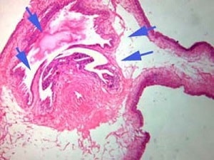
Figure A
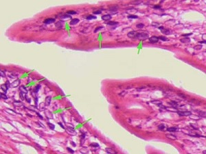
Figure B
More on cysticercosis: https://www.cdc.gov/dpdx/cysticercosis/index.html
Images presented in the dpdx case studies are from specimens submitted for diagnosis or archiving. On rare occasions, clinical histories given may be partly fictitious.
DPDx is an educational resource designed for health professionals and laboratory scientists. For an overview including prevention, control, and treatment visit www.cdc.gov/parasites/.