
Case #435 – January, 2017
An adult patient with right eye and eyelid irritation sought medical attention at an eye clinic. Upon examination, living organisms and what appeared to be egg cases were identified on the patient’s eyelids and eyelashes. A digital photograph (Figure A) was submitted to the DPDx Team for confirmation of the presence of ectoparasites. Specimens were requested by the DPDx Team to allow for a more specific and accurate diagnosis. Submitted were two organisms and a few of the egg cases. Figures B–D, taken with a dissecting microscope, show the organisms that were submitted which measured approximately 1.6 millimeters in length. Figures E and F, taken with a compound microscope at 100x and 200x magnification respectively, show two of the egg cases which averaged 700 micrometers in length. What is your diagnosis? Based on what criteria?
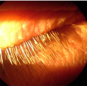
Figure A
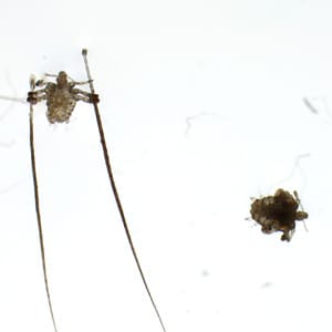
Figure B
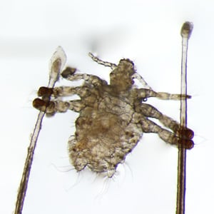
Figure C
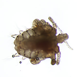
Figure D
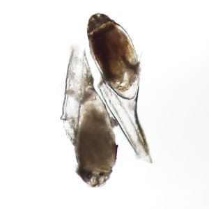
Figure E
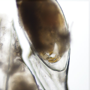
Figure F
This case and Figure A were kindly provided by Solomon Eye Physicians & Surgeons, Bowie, Maryland.
Images presented in the dpdx case studies are from specimens submitted for diagnosis or archiving. On rare occasions, clinical histories given may be partly fictitious.
DPDx is an educational resource designed for health professionals and laboratory scientists. For an overview including prevention, control, and treatment visit www.cdc.gov/parasites/.