
Case #411 – January 2016
A 23-year-old female with no documented travel history presented with iron deficiency anemia and periodic abdominal pain. Ova-and-parasite (O&P) examinations of stool were negative. A colonoscopy was performed and a worm-like object (Figures A and B) was observed attached to the mucosa of the ascending colon. The object was removed, sent to Pathology, and processed by routine histologic work-up. Figures C–E show what was observed by the pathologist after sectioning and staining with hematoxylin-and-eosin (H&E). Images were captures and sent to the CDC-DPDx for diagnostic assistance. What is your diagnosis? Based on what criteria?

Figure A

Figure D

Figure B

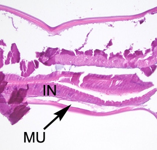
Figure E

Figure C
This case and images were kindly provided by St. David’s South Austin Medical Center, Austin, TX.
DPDx is an educational resource designed for health professionals and laboratory scientists. For an overview including prevention, control, and treatment visit www.cdc.gov/parasites/.