
Case #401 – August 2015
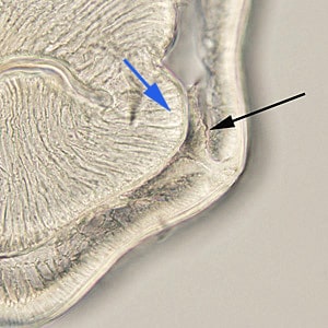
A worm approximately three centimeters long was observed and removed during a routine colonoscopy of a 54-year-old man from Scandinavia. The worm was sent to the CDC-DPDx Team for identification. Figure A shows the gross worm at 10x magnification using a dissecting microscope. It was then cleared with lacto-phenol and examined at 100x magnification (Figures B and C show the anterior and posterior respectively). A thin cross-section was made and examined at the same magnification (Figure D) and 200x (Figure E). What is your diagnosis? Based on what criteria?

Figure A

Figure B

Figure C

Figure D

Figure E
Images presented in the DPDx case studies are from specimens submitted for diagnosis or archiving. On rare occasions, clinical histories given may be partly fictitious.
DPDx is an educational resource designed for health professionals and laboratory scientists. For an overview including prevention, control, and treatment visit www.cdc.gov/parasites/.