
Case #399 – July 2015
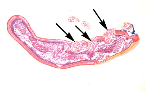
A skin biopsy specimen was collected from the clavicle region of a 45-year-old male who presented with what appeared to be a pigmented lesion. The patient resides in Kentucky and has no known international travel. The biopsy specimen was sent to Pathology for routine histologic work-up. Objects suggestive of an organism were examined on slides stained with hematoxylin-and-eosin (H&E). The attending pathologist captured images and sent them to CDC-DPDx for diagnostic assistance. Figures A–E represent five of the images received. What is your diagnosis? Based on what criteria?

Figure A

Figure B

Figure C

Figure D

Figure E
This case and images were kindly provided by the Lexington Clinic, Lexington, KY.
Images presented in the DPDx case studies are from specimens submitted for diagnosis or archiving. On rare occasions, clinical histories given may be partly fictitious.
DPDx is an educational resource designed for health professionals and laboratory scientists. For an overview including prevention, control, and treatment visit www.cdc.gov/parasites/.