
Case #150 – February, 2005
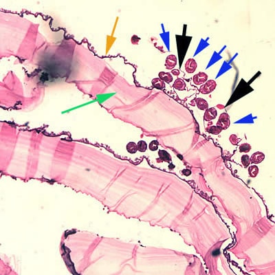
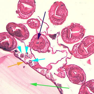
A 28-year-old previously healthy man had a physical examination, including a chest x-ray, as one of his pre-employment requirements. The man had emigrated from Bulgaria approximately one year prior to the examination. The x-ray showed a cyst-like lesion in his right lung. The cyst was later surgically removed, and sent to pathology for identification. The cyst measured 3-4 cm in diameter. The following images were captured from a hematoxylin and eosin (H & E) stained section of the cyst. Figure A was captured at 40× magnification, and Figure B at 200×. What is your diagnosis? Based on what criteria?

Figure A

Figure B
Images presented in the DPDx case studies are from specimens submitted for diagnosis or archiving. On rare occasions, clinical histories given may be partly fictitious.
DPDx is an educational resource designed for health professionals and laboratory scientists. For an overview including prevention, control, and treatment visit www.cdc.gov/parasites/.