bannermenu

Chilomastix mesnili

[Chilomastix mesnili]

Causal Agents

Chilomastix mesnili is a nonpathogenic flagellate that is often described as a commensal organism in the human gastrointestinal tract.

Life Cycle

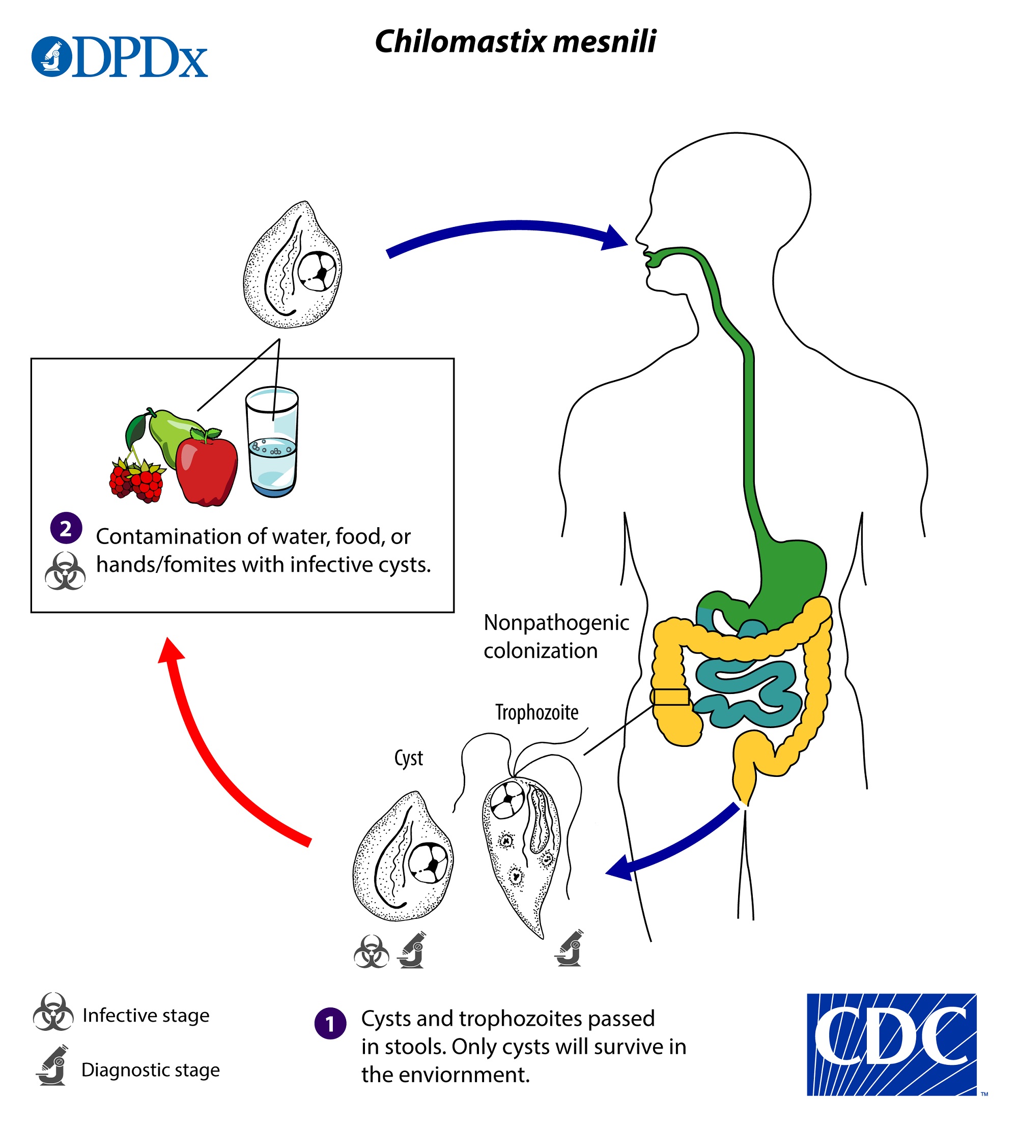
The cyst stage is resistant to environmental pressures and is responsible for transmission of Chilomastix. Both cysts and trophozoites can be found in the feces (diagnostic stages) image . Infection occurs by the ingestion of cysts in contaminated water, food, or by the fecal-oral route (hands or fomites) image . In the large (and possibly small) intestine, excystation releases trophozoites. Chilomastix resides in the cecum and/or colon; it is generally considered a commensal whose contribution to pathogenesis is uncertain.

Geographic Distribution

Worldwide. It is more prevalent in areas with inadequate sanitation.

Clinical Presentation

Chilomastix mesnili is considered nonpathogenic. The presence of cysts and/or trophozoites in stool specimens can however be an indicator of fecal contamination of a food or water source, and thus does not rule out other parasitic infections.

Chilomastix mesnili trophozoites, trichrome stain.


Chilomastix mesnili is a flagellated protozoan generally regarded as nonpathogenic in the human host. Trophozoites are pear-shaped and usually measure 6–24 µm in length. The anterior end is rounded and contains a single nucleus with an eccentric karyosome. The posterior end tapers off to a point.

Figure A: Trophozoite of <em>C. mesnili</em> from a stool specimen, stained with trichrome. Image taken at 1000x magnification.
Figure A: Trophozoite of C. mesnili from a stool specimen, stained with trichrome. Image taken at 1000x magnification.
Figure B: Trophozoite of <em>C. mesnili</em> from a stool specimen, stained with trichrome. Image taken at 1000x magnification.
Figure B: Trophozoite of C. mesnili from a stool specimen, stained with trichrome. Image taken at 1000x magnification.
Figure C: Trophozoite of <em>C. mesnili</em> from a stool specimen, stained with trichrome. Image taken at 1000x magnification.
Figure C: Trophozoite of C. mesnili from a stool specimen, stained with trichrome. Image taken at 1000x magnification.
Figure D: Trophozoite of <em>C. mesnili</em> from a stool specimen, stained with trichrome. Image taken at 1000x magnification.
Figure D: Trophozoite of C. mesnili from a stool specimen, stained with trichrome. Image taken at 1000x magnification.
Chilomastix mesnili cysts, trichrome stain.
Figure A: Cyst of <em>C. mesnili</em> in a stool specimen, stained with trichrome. Image taken at 1000x magnification.
Figure A: Cyst of C. mesnili in a stool specimen, stained with trichrome. Image taken at 1000x magnification.
Figure B: Cyst of <em>C. mesnili</em> in a stool specimen, stained with trichrome. Image taken at 1000x magnification.
Figure B: Cyst of C. mesnili in a stool specimen, stained with trichrome. Image taken at 1000x magnification.
Figure C: Cyst of <em>C. mesnili</em> in a stool specimen, stained with trichrome. Image taken at 1000x magnification.
Figure C: Cyst of C. mesnili in a stool specimen, stained with trichrome. Image taken at 1000x magnification.
Figure D: Cyst of <em>C. mesnili</em> in a stool specimen, stained with trichrome. Image taken at 1000x magnification.
Figure D: Cyst of C. mesnili in a stool specimen, stained with trichrome. Image taken at 1000x magnification.
Figure E: Cyst of <em>C. mesnili</em> in a stool specimen, stained with trichrome. Image taken at 1000x magnification.
Figure E: Cyst of C. mesnili in a stool specimen, stained with trichrome. Image taken at 1000x magnification.
Figure F: Cyst (lower) and trophozoite (upper) of <em>C. mesnili</em> in a stool specimen, stained with trichrome. Image taken at 1000x magnification.
Figure F: Cyst (lower) and trophozoite (upper) of C. mesnili in a stool specimen, stained with trichrome. Image taken at 1000x magnification.
Chilomastix mesnili cysts in wet mounts.
Figure B: Cyst of <em>C. mesnili</em> in a concentrated wet mount of stool, stained with iodine. Image taken at 1000x magnification.
Figure B: Cyst of C. mesnili in a concentrated wet mount of stool, stained with iodine. Image taken at 1000x magnification.

Laboratory Diagnosis

Chilomastix mesnili is identified through the detection of cysts and/or trophozoites in stool specimens, both concentrated wet mounts and permanent stained smears (e.g., trichrome).

Laboratory Safety

Standard precautions for the processing of stool samples apply. While Chilomastix mesnili is non-pathogenic, care should be taken to avoid other enteric pathogens potentially in unfixed stools.

Treatment Information

For information about treatment please contact CDC-INFO.

DPDx is an educational resource designed for health professionals and laboratory scientists. For an overview including prevention, control, and treatment visit www.cdc.gov/parasites/.

Page last reviewed: June 5, 2019