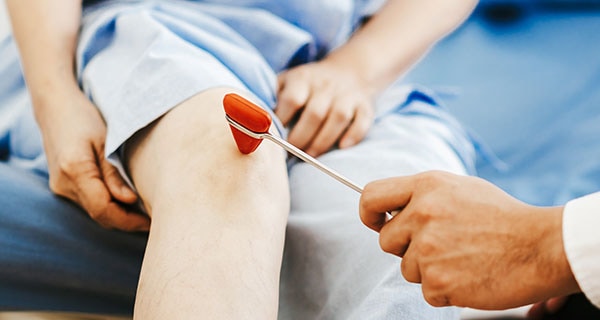
Initial Evaluation and Diagnostic Studies
Initial Evaluation
Medical History
- Collect information on any illness in the past 4 weeks
- Note respiratory and GI symptoms, with or without fever
- Ask about hand, foot, and mouth lesions (possible EV-A71 or similar viral infection)
Focused, Age-appropriate Assessment and Questions to Evaluate Limb Function Impairment(s)
New inability or difficulty
Example questions to ask about limb function
New inability or difficulty
To use arm(s)/hand(s)
Example questions to ask about limb function
- Can they feed themselves?
- Are they suddenly using one limb less or refusing to use one limb?
New inability or difficulty
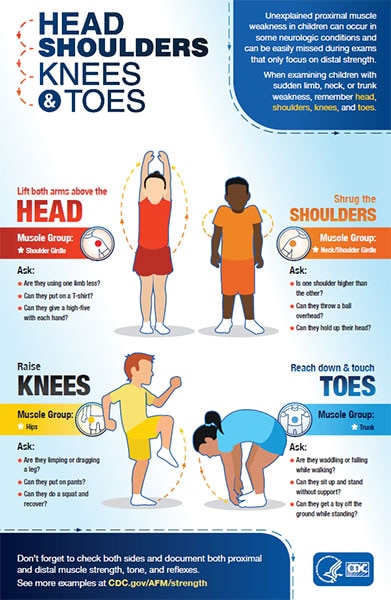
To raise arm(s) above the head
Example questions to ask about limb function
- Can they put on or take off a T-shirt?
- Can they throw a ball overhead?
New inability or difficulty
To walk
Example questions to ask about limb function
- Are they limping or dragging a leg?
- Are they falling often while walking?
New inability or difficulty
To get up unassisted from sitting or squat
Example questions to ask about limb function
- Can they put on or take off pants?
- Can they get out of bathtub unassisted?
Keep in mind that young children or their parents may not describe their limb function impairment as “weakness.”
Additional Signs and Symptoms
Ask about additional signs and symptoms, including:
- Difficulty holding their head up
- Decreased appetite or difficulty swallowing
- Increased sleepiness or inactivity
- Headache or neck, shoulder, or back pain
- Patients often complain of this prior or concurrent to weakness
- Pain in extremities
- Bowel or bladder changes, particularly constipation
Physical Exam
Perform physical exam along with an age-appropriate neurological exam.
Neurological examination should include documentation of:
- Muscle tone (flaccid/loose vs spastic/tight and firm)
- Muscle strength (full strength, move against gravity with some resistance/pressure, move against gravity but with no resistance/pressure, or little limb movement but not against gravity, no muscle movement at all)
- Reflexes in each extremity (hypo-, hyper, or absent)
- Any cranial nerve deficiencies such as for facial, palatal and shoulder asymmetry, hoarseness or hypophonia and dysphagia (if possible)
Note: Sensory exam is often normal in patients with AFM.
Assess the patient’s ability to protect their airway:
- Document respiratory sufficiency
- Negative inspiratory force may be used if the child is old enough and able to cooperate
Check for autonomic manifestations:
- Blood pressure lability
- Body temperature instability
Initial Neurodiagnostic Studies
Laboratory Tests
cell count with differential, protein and glucose; oligoclonal bands; meningitis/encephalitis PCR panel
EV PCR, anti-MOG (myelin oligodendrocyte glycoprotein) and anti-aquaporin antibodies, HSV, EBV, WNV
EV PCR
Respiratory multiplex testing and enterovirus (EV) PCR
MRI of the spine and brain
Consider additional pathogen-specific testing (e.g., Lyme) based on seasonality, exposures, and geography.
Note: Some of these studies may require sedation, depending on child’s age
- Coordinate the procedures to avoid repetitive sedation if possible
- Monitor respiratory status continuously
Reference: Hardy D, Hopkins SE. Update on AFM: Recognition, reporting, aetiology, and outcomesexternal icon. Arch Dis Child 2020. DOI: 10.1136/archdischild-2019-316817. Online ahead of print.
Laboratory Specimen Collection
Rapid specimen collection increases the chance of pathogen detection. Specific testing for AFM should be done in consultation with a neurologist and infectious disease specialists. Consider additional pathogen-specific testing based on seasonality, exposures, and geography and clinical presentation.
CSF, respiratory (NP/OP), serum, and stool specimens should be also sent to CDC for surveillance testing. Contact your health department to coordinate sending of specimens to CDC for testing.
MRI Imaging
Order MRI of the spine and brain with and without contrast.
- Use the highest tesla scanner available (ideally 3T).
Imaging within the first 72 hours of limb weakness may be normal and should be repeated if clinically indicated.
- Axial and sagittal images are most helpful in identifying lesions.
- Multiple levels of the spinal cord are often involved, consider imaging entire spinal cord.
- In patients with cranial nerve deficits, high cuts of brainstem or total brain MRI should be considered.
- Although lesions are predominantly gray matter, some patients with AFM may also have some white matter involvement.
Reference: Maloney JA et al. MRI Findings in Children With Acute Flaccid Paralysis and Cranial Nerve Dysfunction Occurring During the 2014 Enterovirus D68 Outbreakexternal icon. Am J Neuroradiol 2015;36(2):245-50.