Case #486 – February, 2019
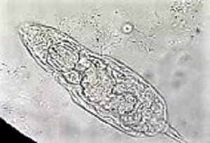
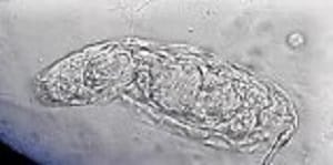
A 10-year-old female patient from Argentina was taken to the hospital by her parent with complaints of “not feeling well.” A urine sample was collected and examined as part of the routine checkup. Microscopic examination of the urine sediment using the 40x objective lens revealed the following: 0-1 WBC and 0-2 RBC per field and a motile organism shown in the video that was sent to the DPDx team for identification. Still images of the organism were made by capturing screenshots at different points of the video (Figures A – C). What is your diagnosis? Based on what criteria?
This case and video was kindly provided by the Hospital Clinicas de Cordoba/Universidad de Cordoba, Argentina.

Figure A

Figure B

Figure C
Images presented in the dpdx case studies are from specimens submitted for diagnosis or archiving. On rare occasions, clinical histories given may be partly fictitious.
DPDx is an educational resource designed for health professionals and laboratory scientists. For an overview including prevention, control, and treatment visit www.cdc.gov/parasites/.