Case #481 – December, 2018
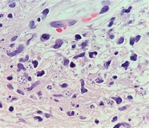
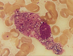
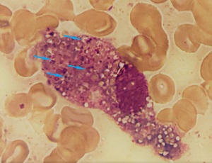
A 22-year-old male noticed painless, ulcerating lesions with crusting and mild swelling on his back and face. He had travelled to the rain forest of Peru five months earlier, where he experienced multiple insect bites all over his body. A biopsy specimen from a lesion on his back was sectioned and stained with hematoxylin and eosin (H&E) (Figure A). A smear of a scraping from the biopsy site was also made and stained with Giemsa stain (Figure B). Both figures show what was observed on the slides.
What is your diagnosis? Based on what criteria? What other testing would you recommend?
This case and images were kindly provided by the Pathology Department, Stony Brook University Hospital, Stony Brook, NY.

Figure A

Figure B
Images presented in the dpdx case studies are from specimens submitted for diagnosis or archiving. On rare occasions, clinical histories given may be partly fictitious.
DPDx is an educational resource designed for health professionals and laboratory scientists. For an overview including prevention, control, and treatment visit www.cdc.gov/parasites/.

