Case #459 – January, 2018
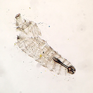
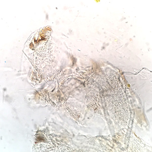
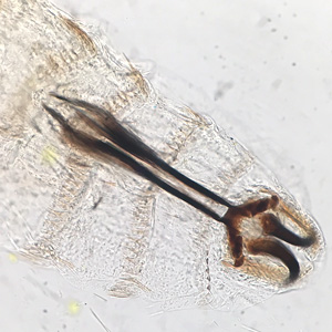
A visitor to Hawaii experienced onset of moderate irritation of one eye accompanied by redness and the sensation of a foreign body. The visitor removed a few small, 1-2 millimeter long white-colored motile organisms from the conjunctiva of the affected eye. The organisms were placed on the end of a tube of lip balm for transport to a health care facility for identification. Figures A–C are images taken with the camera of a cellular phone via the eyepiece of a microscope and show details of one of the organisms. What is your diagnosis? Based on what criteria?
This case and images were kindly provided by ARUP Laboratories and The University of Utah School of Medicine, Infectious Diseases Division, both in Salt Lake City, Utah.

Figure A

Figure B

Figure C
Images presented in the dpdx case studies are from specimens submitted for diagnosis or archiving. On rare occasions, clinical histories given may be partly fictitious.
DPDx is an educational resource designed for health professionals and laboratory scientists. For an overview including prevention, control, and treatment visit www.cdc.gov/parasites/.