
Case #457 – December, 2017
A nine-year-old female refugee from Eritrea with neutropenia and eosinophil counts within normal range had a stool examination as part of a refugee screening. Microscopic examination of an iodine wet mount preparation of a formalin-ethyl acetate (FEA) concentration of the stool sample submitted found what is shown in Figures A and B (taken at 400x) and C (at 1000x). What is your diagnosis? Based on what morphologic features?
This case and images were by provided by the Cadham Provincial Public Health Laboratory, Winnipeg, MB.

Figure A

Figure B

Figure C
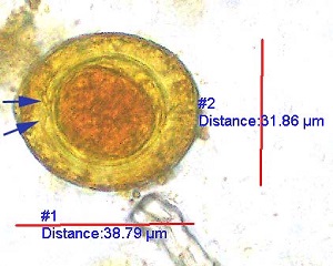
Figure A
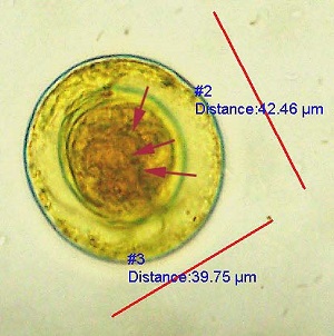
Figure B
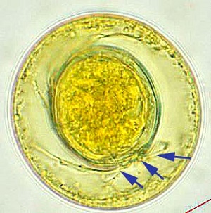
Figure C
This was a case of hymenolepiasis caused by Hymenolepis (Rodentolepis) nana (the dwarf tapeworm). Morphologic features shown included:
- Eggs within the size range for H. nana (30-50 micrometers).
- Presence of an oncosphere with hooklets, which are somewhat inconspicuous in image (red arrows; Figures B).
- Polar filaments between the inner and outer membranes (blue arrows, Figure A and C).
Infection has worldwide distribution and is most commonly seen in children
More on hymenolepiasis: https://www.cdc.gov/dpdx/hymenolepiasis/index.html
Images presented in the dpdx case studies are from specimens submitted for diagnosis or archiving. On rare occasions, clinical histories given may be partly fictitious.
DPDx is an educational resource designed for health professionals and laboratory scientists. For an overview including prevention, control, and treatment visit www.cdc.gov/parasites/.