
Case #454 – October, 2017
An eleven-year-old male from Arizona with no known past medical history was evaluated in the ophthalmology clinic after a 3-4 week history of progressive redness in his left eye, with associated pain and growth of a nodule. There was no record of travel outside the US. However, the patient went on a trip with his school on the Colorado River. An excisional biopsy of the left eye conjunctiva revealed objects consistent with a parasitic worm infection. Figures A – E show what was found in the hematoxylin & eosin (H&E) stain of the biopsy. What is your diagnosis? Based on what morphologic features?
This case was kindly contributed by the Arizona State Health Department

Figure A

Figure B

Figure C

Figure D

Figure E
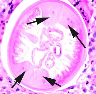
Figure C
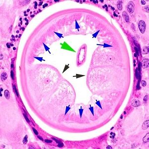
Figure D

Figure E
This was a case of zoonotic onchocerciasis caused by Onchocerca lupi. Diagnostic features included:
- Prominent lateral cords Figures C and D (black arrows)
- Short coelomyarian muscle cells Figure D (blue arrow)
- Intestine Figure D (green arrow)
- Striae Figure E, (green arrows) that run between and below the cuticular ridges Figure E (black arrows)
Images presented in the dpdx case studies are from specimens submitted for diagnosis or archiving. On rare occasions, clinical histories given may be partly fictitious.
DPDx is an educational resource designed for health professionals and laboratory scientists. For an overview including prevention, control, and treatment visit www.cdc.gov/parasites/.