Case #437 – February, 2017
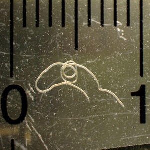
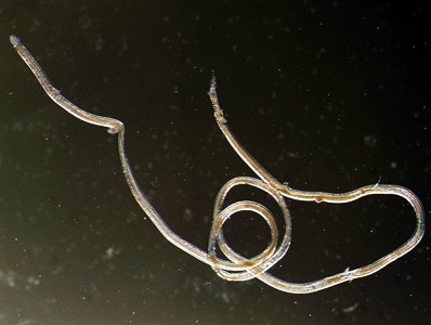
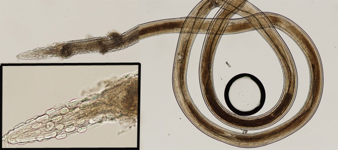
A 31-year-old female, with travel to Mexico a few months prior, noticed a small worm-like object on the inner surface of her lower lip. She removed the object with a needle and delivered it to her health care provider. The object was forwarded to the CDC DPDx Team for identification. Figures A and B were taken with a dissecting microscope and the ruler in the background shows one centimeter; Figure C is a composite of two images of the organism that was submitted taken with a compound microscope to show the entire organism at 40x magnification. The insert (of the anterior end) was taken at 100x magnification. What is your diagnosis? Based on what criteria?

Figure A

Figure B

Figure C
This case was kindly provided by Optimus Medical Clinic, Houston, Texas.
Images presented in the dpdx case studies are from specimens submitted for diagnosis or archiving. On rare occasions, clinical histories given may be partly fictitious.
DPDx is an educational resource designed for health professionals and laboratory scientists. For an overview including prevention, control, and treatment visit www.cdc.gov/parasites/.