
Case #384 – November 2014
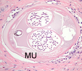
A 73-year-old woman presented to a local clinic in Ghana with a skin nodule that had developed adjacent to the resection site of a previous low-grade malignant skin tumor. Biopsy specimens were collected and sent to a lab in the U.S. for histologic processing, including sectioning and staining with hematoxylin-and-eosin (H&E). Images of suspect structures were captured and sent to the DPDx Team for diagnostic assistance. Figures A–E show several of the images that were received for analysis. Neither sizes nor magnifications were provided with the images. What is your diagnosis? Based on what criteria?

Figure A

Figure B

Figure C

Figure D

Figure E
This case and images were kindly provided by Saint Joseph Mercy Hospital, Ann Arbor, MI.
Images presented in the DPDx case studies are from specimens submitted for diagnosis or archiving. On rare occasions, clinical histories given may be partly fictitious.
DPDx is an educational resource designed for health professionals and laboratory scientists. For an overview including prevention, control, and treatment visit www.cdc.gov/parasites/.