Case #345 – April, 2013
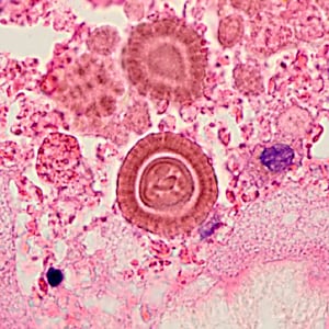
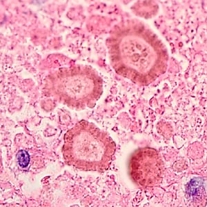
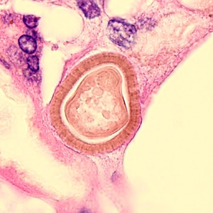
An 83-year-old man presented to a medical institution with shortness of breath and left flank pain that developed during a trip to Mexico shortly beforehand. Magnetic resolution imaging (MRI) revealed an exophytic mass on his left kidney. Approximately 4 months later he underwent surgery and the mass was sent to Pathology for analysis. The pathology report stated foreign elements of unknown etiology observed in perinephritic fat. Gomori-Grocott methenamine silver (GMS) and periodic acid-Schiff (PAS) stains were negative for fungal organisms. Images were captured of the objects of interest and sent to DPDx for consult. Figures A–C show the objects; neither magnification used nor size of the objects was given. What is your diagnosis? Based on what criteria?

Figure A

Figure B

Figure C
this case and images were kindly provided by the David Geffen School of Medicine at UCLA, Los Angeles, CA.
Images presented in the DPDx case studies are from specimens submitted for diagnosis or archiving. On rare occasions, clinical histories given may be partly fictitious.
DPDx is an educational resource designed for health professionals and laboratory scientists. For an overview including prevention, control, and treatment visit www.cdc.gov/parasites/.