Case #316 – January, 2012
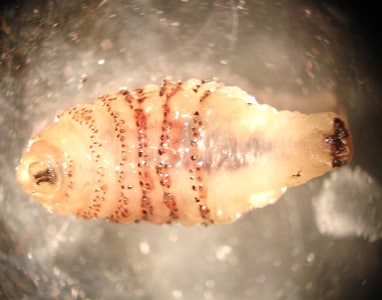
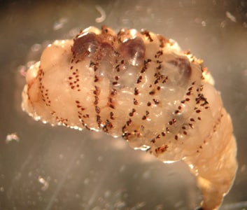
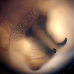
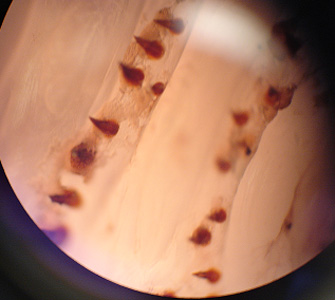
A 75-year-old man developed a lesion on his right forearm. The lesion developed approximately one month after returning from a trip to Costa Rica. The patient’s attending physician removed what appeared to be a fly larva. The larva was sent to the hospital’s Microbiology Department for further identification. Figure A and B show the ventral and lateral sides of the larva, respectively. Figure C shows a close-up of the mouthparts. Figure D shows a close-up of the cuticular spines. What is your identification? Based on what criteria?

Figure A

Figure B

Figure C

Figure D
This case and images were kindly provided by HealthEast St. Joseph’s Hospital, St. Paul, MN.
Images presented in the DPDx case studies are from specimens submitted for diagnosis or archiving. On rare occasions, clinical histories given may be partly fictitious.
DPDx is an educational resource designed for health professionals and laboratory scientists. For an overview including prevention, control, and treatment visit www.cdc.gov/parasites/.