
Case #288 – November, 2010
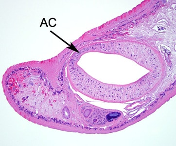
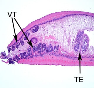
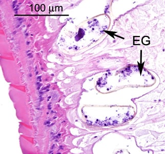
A 50-year-old man underwent a screening colonoscopy after complaints of constipation. A polyp measuring approximately 3 mm was observed during the procedure. A biopsy of the polyp was taken and sent to a pathology laboratory for sectioning and staining. Figures A–D show what was observed by the attending pathologist. The image in Figure A was captured at 40x magnification. The images in Figures B and C were captured at 100x magnification. The image in Figure D was captured at 400x magnification. What is your diagnosis? Based on what criteria?

Figure A

Figure B

Figure C

Figure D
This case, and the specimen from which images were taken, were courtesy of the New York State Health Department, Wadsworth Center.
Images presented in the DPDx case studies are from specimens submitted for diagnosis or archiving. On rare occasions, clinical histories given may be partly fictitious.
DPDx is an educational resource designed for health professionals and laboratory scientists. For an overview including prevention, control, and treatment visit www.cdc.gov/parasites/.