
Case #278 – June, 2010
DPDx is moving! Along with the all of the laboratories within the Division of Parasitic Diseases and Malaria at the CDC! We want to take this opportunity to let you know that although actual specimen receiving and testing will be temporarily interrupted for about a week or two, DPDx will continue to provide telediagnosis assistance via the internet.
This leads us to the second case for this month. While packing for the move, we discovered an interesting pathology slide labeled with only an accession number. Figure A shows the slide and Figure B shows the specimen using a dissecting microscope. The slide was observed under a compound microscope for higher magnification; Figures C and E show areas captured at 40x, and Figures D and F at 200x. What is your diagnosis? Based on what criteria?
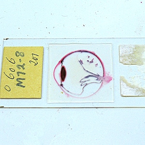
Figure A
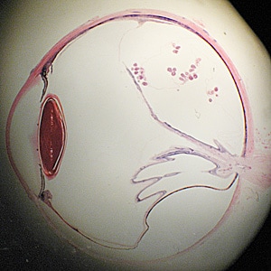
Figure B
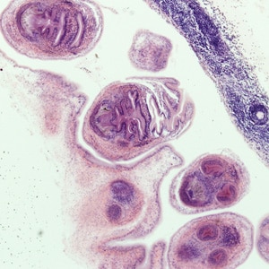
Figure C
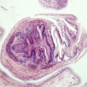
Figure D
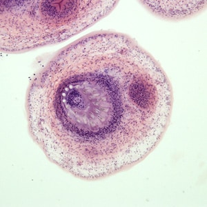
Figure E
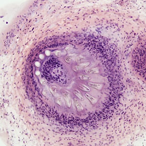
Figure F
Images presented in the DPDx case studies are from specimens submitted for diagnosis or archiving. On rare occasions, clinical histories given may be partly fictitious.
DPDx is an educational resource designed for health professionals and laboratory scientists. For an overview including prevention, control, and treatment visit www.cdc.gov/parasites/.