
Case #275 – May, 2010
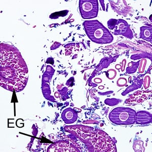
A 70-year-old female, who had recently returned from a trip to Madagascar, went to the hospital for a painful sensation on the underside of her left foot while walking. Examination of the area between the hallux and index toes revealed an ulcerative lesion. A biopsy was performed and sent to the Pathology Department for work-up. The specimen was sectioned, stained with hematoxylin and eosin (H&E) and examined by the attending pathologist. Figures A and B show what was observed at 40x magnification. Figures C and D show the same fields at 200x magnification, respectively. What is your diagnosis? Based on what criteria?

Figure A

Figure B

Figure C

Figure D

Images presented in the DPDx case studies are from specimens submitted for diagnosis or archiving. On rare occasions, clinical histories given may be partly fictitious.
DPDx is an educational resource designed for health professionals and laboratory scientists. For an overview including prevention, control, and treatment visit www.cdc.gov/parasites/.