
Case #186 – August, 2006
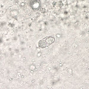
A man went to a local hospital with abdominal pain and weight loss. He reported that he frequently travels to South America and had previously been diagnosed with ascariasis, although recent stool specimens were negative. The laboratory saw the objects in Figures A, B, and C in the patient’s stool samples. The objects measured 10 to 15 µm. Figure A was taken at 400× magnification from wet mount stained with iodine. Figures B and C were taken at 1000× magnification from a trichrome stained slide. What is your diagnosis? Based on what criteria?

Figure A

Figure B

Figure C
This case was kindly contributed by the Wisconsin State Laboratory of Hygiene.
Images presented in the DPDx case studies are from specimens submitted for diagnosis or archiving. On rare occasions, clinical histories given may be partly fictitious.
DPDx is an educational resource designed for health professionals and laboratory scientists. For an overview including prevention, control, and treatment visit www.cdc.gov/parasites/.