
Case #176 – March, 2006
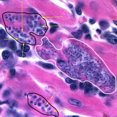
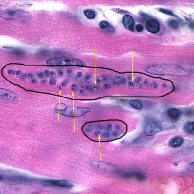
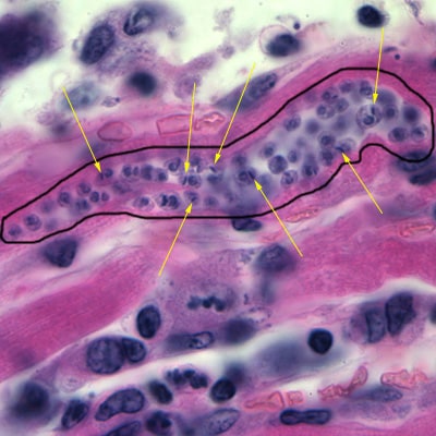
A CDC microbiologist was looking through slide boxes that had been archived to determine whether the slide quality was good enough to continue storing them. He came across a pathology slide of heart tissue that was dated “1929.” Figures A-C show what was observed on the H & E stained preparation. What objects do you see in the images?

Figure A

Figure B

Figure C
This case was kindly contributed by the Auckland City Hospital, New Zealand and images were taken by Dr. Allen Heath from AgResearch-Wallaceville Animal Research Centre.
Images presented in the DPDx case studies are from specimens submitted for diagnosis or archiving. On rare occasions, clinical histories given may be partly fictitious.
DPDx is an educational resource designed for health professionals and laboratory scientists. For an overview including prevention, control, and treatment visit www.cdc.gov/parasites/.