
Case #172 – January 2006
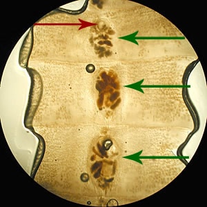
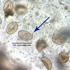
A 45-year-old female noticed a long, worm-like object in her stool. The object was collected and, along with tissue sections prepared by the hospital where she was seen, submitted to the Florida public health laboratory for examination. The specimens were forwarded to CDC’s parasitology diagnostic reference laboratory for identification. Examination methods were: whole worm was examined for morphologic features (Figure A); a portion of the worm was removed (Figure B), cleared using lacto-phenol solution, flattened using two 2″ by 3″ glass slides, and examined under a dissecting microscope (Figure C); and the proglottid was ruptured to release some of the eggs and examined with a compound microscope. Images of the eggs were captured at 40× (Figure D), 100× (Figure E), and 200× (Figure F) magnification. What is your identification? Based on what criteria? Would you recommend any additional confirmatory diagnostic testing?

Figure A

Figure B

Figure C

Figure D

Figure E

Figure F
This case was kindly contributed by the Florida Department of Health.
Images presented in the DPDx case studies are from specimens submitted for diagnosis or archiving. On rare occasions, clinical histories given may be partly fictitious.
DPDx is an educational resource designed for health professionals and laboratory scientists. For an overview including prevention, control, and treatment visit www.cdc.gov/parasites/.