Error processing SSI file
Case #168 – November, 2005
In April 2005, DPDx telediagnosis inquiries received a request for assistance from the Oregon State Public Health Laboratory. Images from a thick and thin blood smear were submitted. Based on information that came with the specimen, the smears were most likely stained with Giemsa. The only case history available was that the patient lived in Africa until the beginning of this year. Figures A-C show what was observed on the thick smear. Figure C is a close-up of the posterior end of the object in Figure B. Figures D-F show what was observed on the thin smear. In Figure D, the object measured 300 micrometers; close-ups of the posterior and anterior ends are shown in Figures E and F, respectively. What is your diagnosis? Based on what criteria?
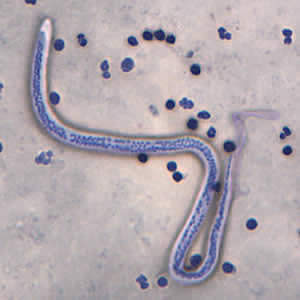
Figure A
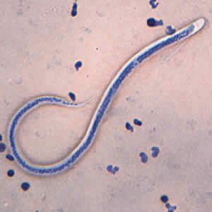
Figure B
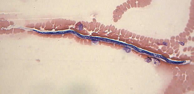
Figure C
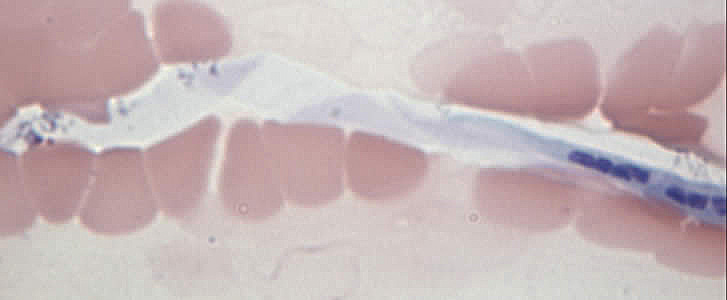
Figure D

Figure E
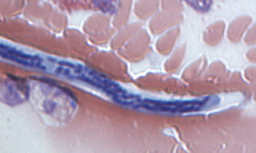
Figure F
This case was kindly contributed by the Oregon State Public Health Laboratory.
Images presented in the DPDx case studies are from specimens submitted for diagnosis or archiving. On rare occasions, clinical histories given may be partly fictitious.
Error processing SSI file