
Case #131 - May, 2004
A 47-year-old man, originally from Thailand, presented to the hospital with upper abdominal pain accompanied with liver enlargement. A biopsy of the bile duct revealed fibrotic thickening. The biopsy specimen was sent to Pathology for routine sectioning and staining. Figures A–F show what was observed by the attending pathologist on slides stained with hematoxylin-and-eosin (H&E). Figures A and B were taken at 100x magnification. Figures C and D were taken at 400x magnification. Figures E and F were taken at 1000x magnification with oil. What is your diagnosis? Based on what criteria?
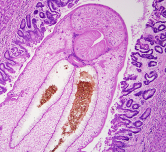
Figure A
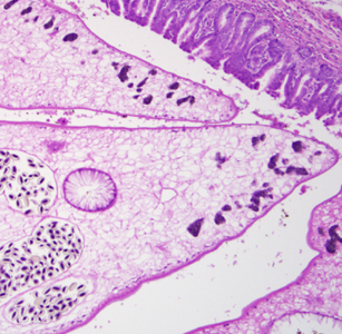
Figure B
Figure C
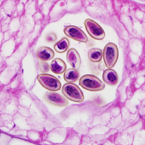
Figure D

Figure E
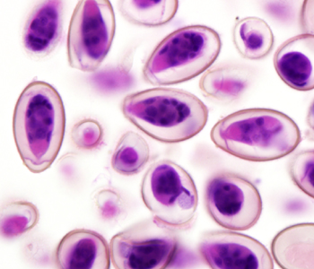
Figure F
This was a case of clonorchiasis caused by the liver fluke, Clonorchis sinensis. Based on the images and clinical presentation in this case, it was not possible to distinguish between C. sinensis and Opisthorchis spp., and opisthorchiasis would have also been an acceptable diagnosis. Diagnostic morphologic features included:
- several characteristics of adult trematodes, including the presence of an oral sucker (OS, Figure A), a thick, muscled pharynx (PH, Figure A), a branching intestinal cecum (CE, Figure A), a uterus filled with eggs (UT, Figure B) and vitteline glands (VT, Figure B).
- the presence of eggs that were within the size range for C. sinensis that possessed an operculum (OP, Figure E) and abopercular knob (KN, Figure E).
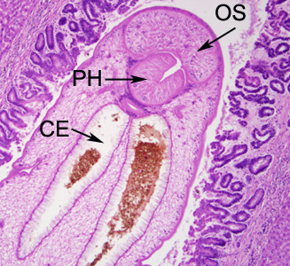
Figure A
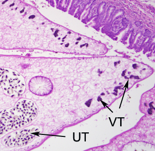
Figure B
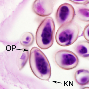
Figure E
More on: Clonorchiasis
Images presented in the monthly case studies are from specimens submitted for diagnosis or archiving. On rare occasions, clinical histories given may be partly fictitious.
DPDx is an educational resource designed for health professionals and laboratory scientists. For an overview including prevention, control, and treatment visit www.cdc.gov/parasites/.