
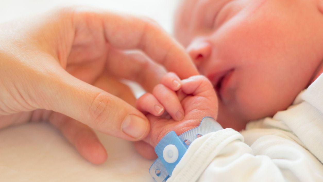
Learn more about congenital heart defects (CHDs) and the different types of conditions.
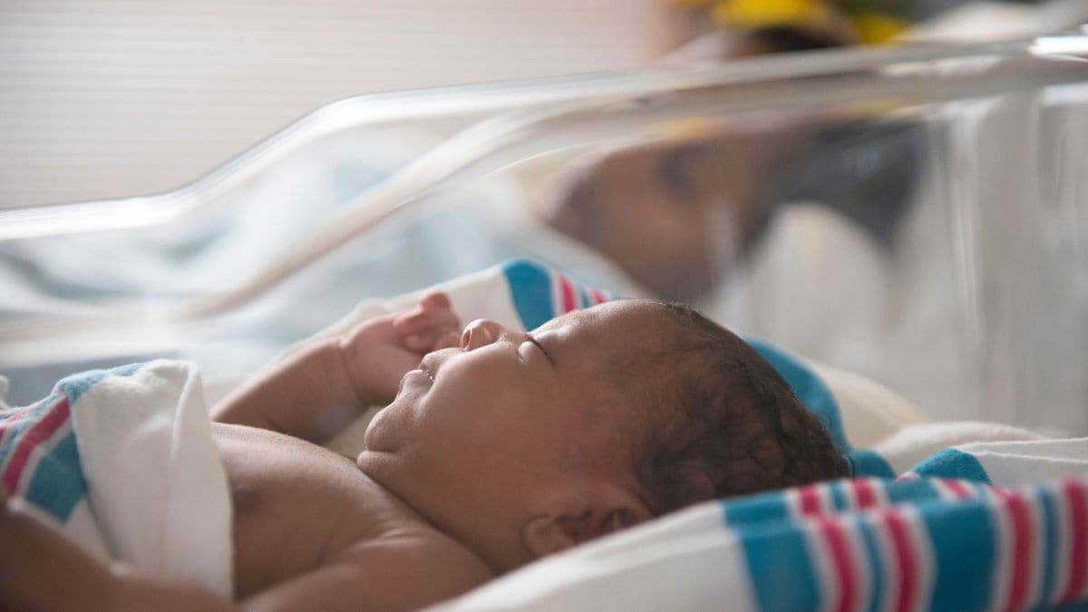
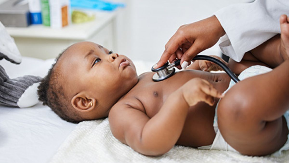
Newborn screening can help identify a critical CHD, so babies can receive prompt care and treatment.
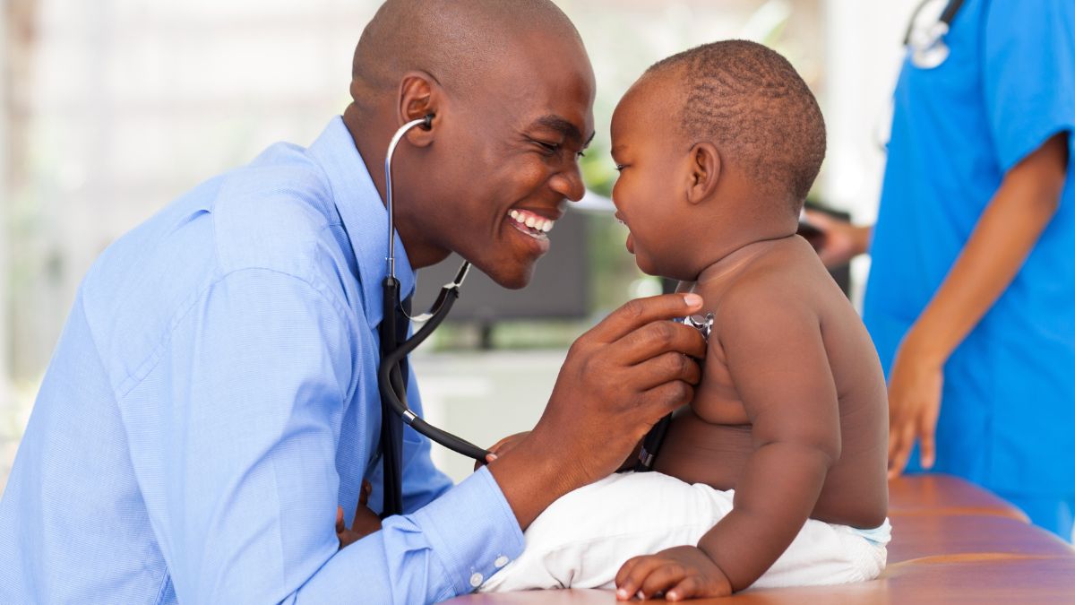
People with heart defects are living longer and healthier lives thanks to better care.

Use these resources to help raise awareness about CHDs and "Heart Heroes."
Living with CHD
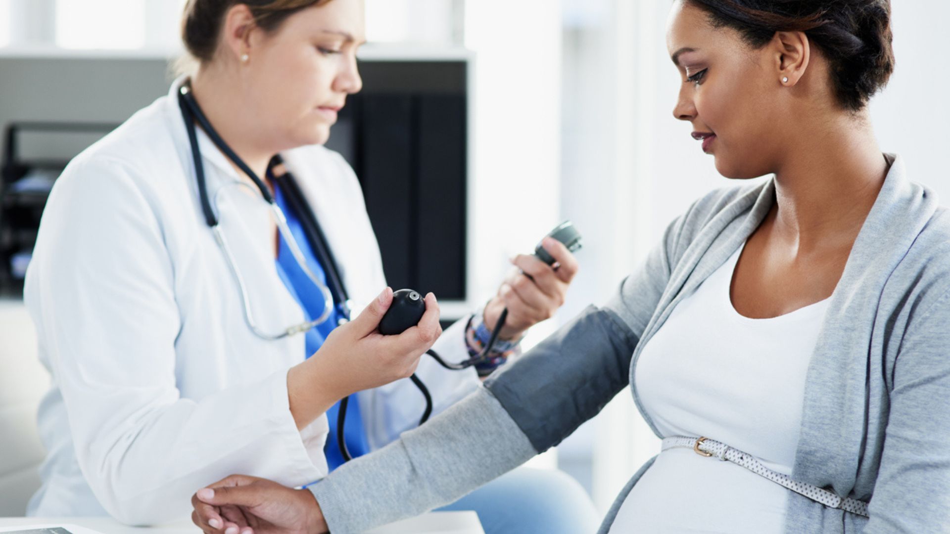
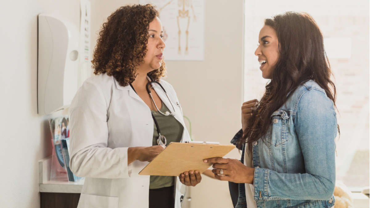
Women living with a heart defect face unique reproductive health issues.
People with CHDs need to have a plan to move from pediatric to adult health care.
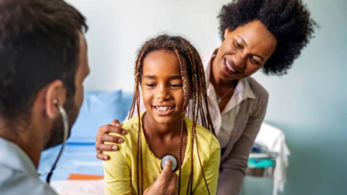
People with heart defects can develop other health issues related to their condition over time.
More Information on CHDs

See how your heart works and moves blood to every part of your body.

Video commentary, podcasts, and webinars that talk about heart defects.
View recent data and statistics on individuals living with heart defects.
Heart defects tracking and research helps us understand causes and improve health of those affected.

CHD toolkit helping physicians promote lifelong cardiac care for people born with heart defects.
Screening for CCHDs helps identify some babies with a CCHD before going home from the hospital.