
Case #134 - June, 2004
A 42-year-old man presented to his health care provider with fever, chills, and mild myalgia. He mentioned that his occupation requires frequent travel to Indonesia and Malaysia and that he often does not take any malaria prophylaxis. Blood smears were ordered, stained with Giemsa, and examined. Figures A–D show what was observed on thick smears at 1000x magnification with oil. Figures E–I show what was observed on thin smears at 1000x magnification with oil. What is your diagnosis? Based on what criteria?
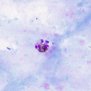
Figure A
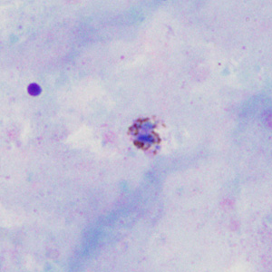
Figure B
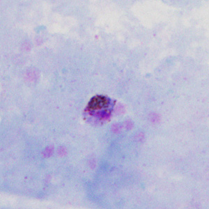
Figure C
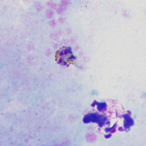
Figure D
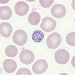
Figure E
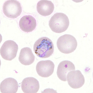
Figure F
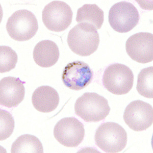
Figure G
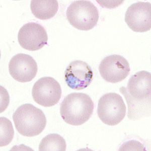
Figure H
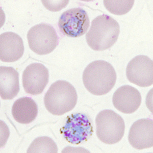
Figure I
Images presented in the monthly case studies are from specimens submitted for diagnosis or archiving. On rare occasions, clinical histories given may be partly fictitious.
DPDx is an educational resource designed for health professionals and laboratory scientists. For an overview including prevention, control, and treatment visit www.cdc.gov/parasites/.